
System and method for magnetically concentrating and detecting biomarkers

a magnetically concentrated and biomarker technology, applied in the field of magnetically concentrated and detecting biomarkers, can solve the problems of limited biomarker repertoire, limited range of biomarkers that are detectable by immunochromatography, and limited sensitivity, so as to achieve the effect of increasing the sample volum

Inactive Publication Date: 2013-04-23
UNIV OF WASHINGTON
View PDF143 Cites 18 Cited by
  • Summary
  • Abstract
  • Description
  • Claims
  • Application Information

AI Technical Summary

Benefits of technology

This approach enhances the sensitivity of diagnostic tests by enabling the detection of biomarkers from diluted samples, overcoming the limitations of existing technologies and improving the range of detectable biomarkers, while maintaining simplicity and portability.

Problems solved by technology

However, these devices suffer from a limitation in sensitivity related to the small sample volume that limits the biomarker repertoire to those at relatively higher blood and plasma concentrations.
However, as noted above, the range of biomarkers that are detectable by immunochromatography is ultimately limited to proteins of relatively high abundance in the plasma in part because of the inability of an immunochromatographic flow strip to process larger volumes of biological sample so as to accumulate sufficient quantities of dilute labeled biomarkers to generate a detectable signal.

Method used


Image

  • System and method for magnetically concentrating and detecting biomarkers
  • System and method for magnetically concentrating and detecting biomarkers
  • System and method for magnetically concentrating and detecting biomarkers

Examples


example 1

Preparation and Characterization of Representative Magnetic and Non-Magnetic Particles and Their Use in a Representative Method of the Invention

[0171]In this example, the preparation and characterization of representative magnetic and non-magnetic particles useful in the methods of the invention are described. The use of these particles in a representative method of the invention is also described.

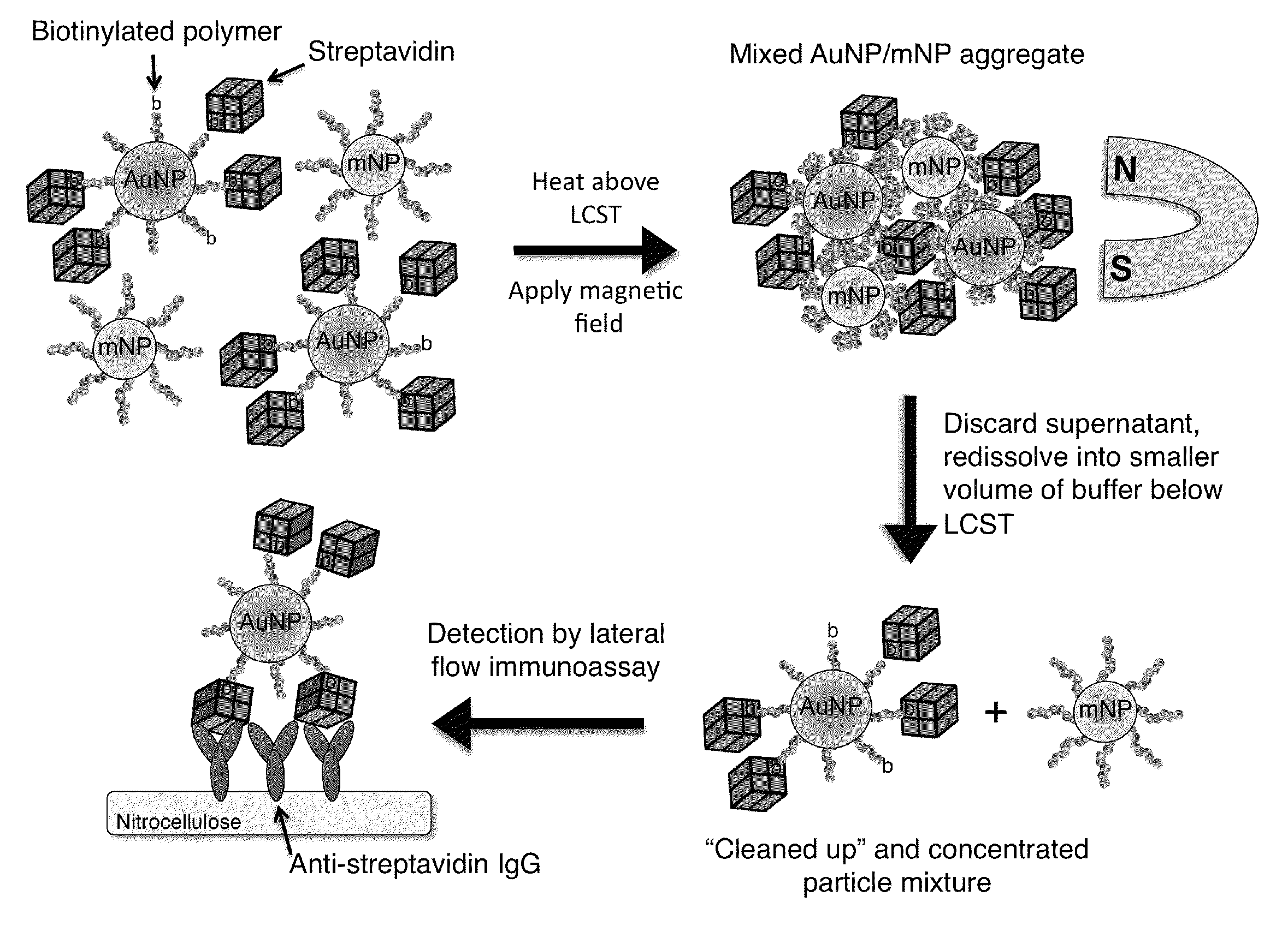
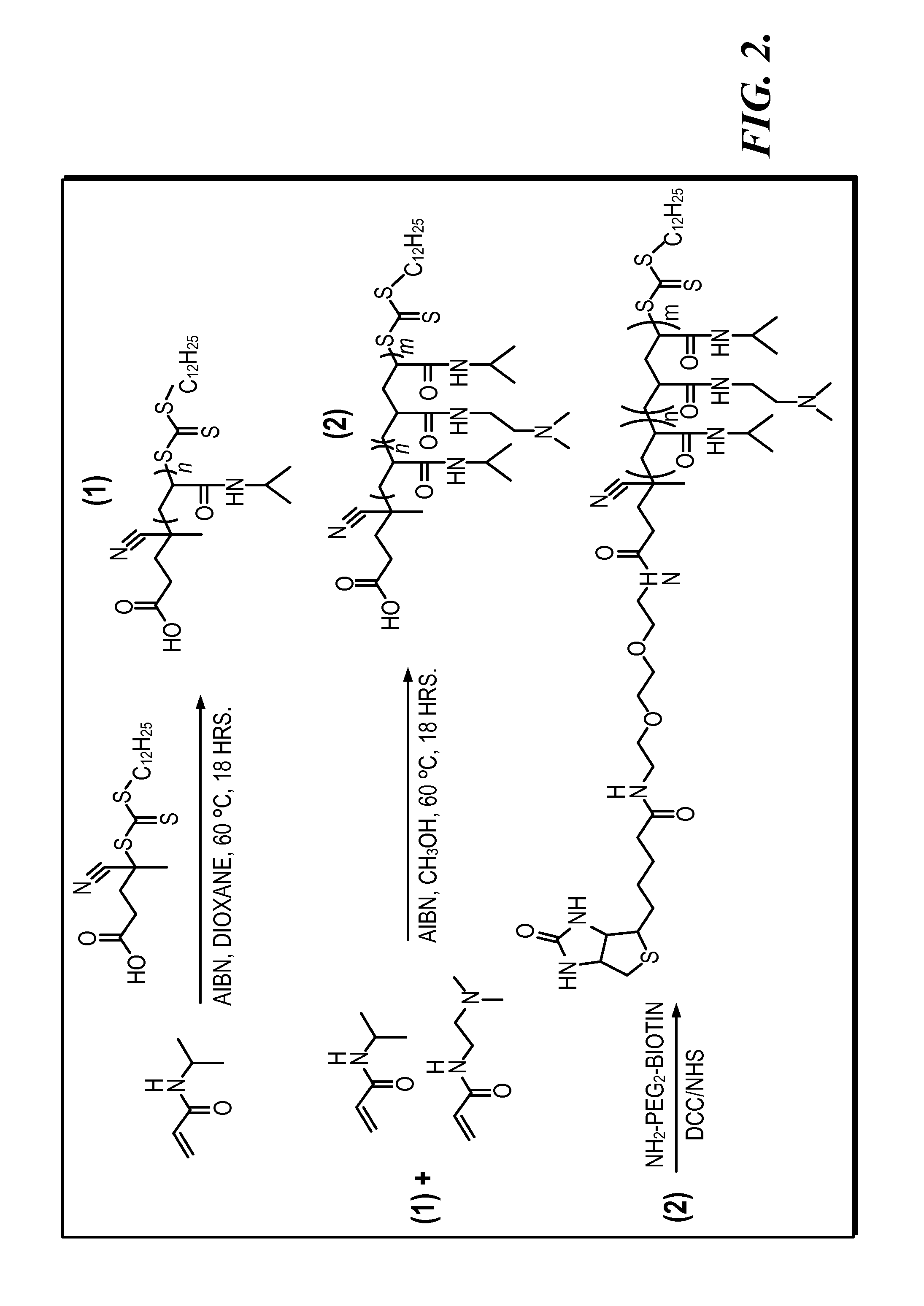
[0172]System Design. The bioseparation / enrichment system consists of a mixture of magnetic and gold nanoparticles, each with a “smart” polymer coating. Gold nanoparticles (about 25 nm diameter) were modified with a biotinylated amine-containing diblock copolymer, and magnetic nanoparticles (about 10 nm diameter) were synthesized directly with a homo-pNIPAAm polymer surface coating. When mixtures of these two particle types were heated above the polymer LCST, the particles co-aggregated driven by hydrophobic interactions between the collapsed polymers. 8 kDa homo-pNIPAAm (2 mg / mL) was added...

example 2

Preparation and Characterization of Representative Magnetic and Non-Magnetic Particles and Their Use in a Representative Multiplexed Method of the Invention

[0196]In this example, the preparation and characterization of representative magnetic and non-magnetic particles useful in the methods of the invention are described. The use of these particles in a representative multiplexed method of the invention is also described.

[0197]FIG. 9 is a schematic illustration of a representative non-magnetic particle useful in the practice of the methods of the invention for capturing a biomarker. This universal bioconjugate design allowed for facile multiplexing by simply adding multiple different biotinylated antibodies to the sample before addition of the universal streptavidin-gold reagent. FIG. 10 is a schematic representation of a representative method of the invention, a magnetic enrichment lateral flow immunoassay, utilizing the universal streptavidin-gold reagent.

[0198]Polymer Synthesis. ...


PUM

PropertyMeasurementUnit
volumeaaaaaaaaaa
concentrationaaaaaaaaaa
volumesaaaaaaaaaa

Abstract

System and method for capturing, concentrating, and detecting a diagnostic target in a liquid, comprising applying a magnetic field to a mixture comprising a co-aggregate in the liquid to provide a collected co-aggregate in the liquid, wherein the co-aggregate comprises a magnetic particle having a stimuli-responsive polymer attached thereto and a non-magnetic particle having a stimuli-responsive polymer and a diagnostic target attached thereto.

Description

CROSS-REFERENCES TO RELATED APPLICATIONS[0001]This application claims the benefit of U.S. Provisional Application No. 61 / 186,780, filed Jun. 12, 2009, and U.S. Provisional Application No. 61 / 259,545, filed Nov. 9, 2009, each incorporated herein by reference in its entirety.STATEMENT OF GOVERNMENT LICENSE RIGHTS[0002]This invention was made with Government support under Grant No. EB000252 awarded by National Institutes of Health. The Government has certain rights in the invention.BACKGROUND OF THE INVENTION[0003]Diagnostic testing is expanding from centralized hospital labs to more distributed settings in both the developed and developing worlds. A current dominant point-of-care diagnostic test format is the lateral flow immunoassay (LFIA), which relies on capillary wicking of fluids through porous nitrocellulose strips. Such tests typically rely on the specific binding between gold-labeled biomarkers with capture antibodies immobilized at the test line of the solid phase. Current de...

Claims


Application Information

Patent Timeline
Patent Type & Authority Patents(United States)
IPC IPC(8): C12M1/00G01N31/22G01N9/00G01N31/00
CPCG01N33/558Y10T436/14Y10T436/143333Y10T436/142222G01N33/54333
Inventor STAYTON, PATRICK S.NASH, MICHAELLAI, JRIUAN
Owner UNIV OF WASHINGTON
Learn More