
Transcorporeal spinal decompression and repair system and related method

a transcorporeal spinal and repair system technology, applied in the field of transcorporeal spinal decompression and repair system and related methods, can solve the problems of loss of function, long recovery time, inherent problems of fusion and disc replacement,

Active Publication Date: 2012-12-04
GLOBUS MEDICAL INC
View PDF150 Cites 21 Cited by
  • Summary
  • Abstract
  • Description
  • Claims
  • Application Information

AI Technical Summary

Problems solved by technology

The fusion of the vertebral segments, however, can lead to problematic results.
In spite of the benefits that these procedures have brought to patients, both fusion and disc replacement have inherent problems.
The surgeries are extensive, recovery time is relatively long, and there is often a loss of function, particularly with the use of fusion implants.
The long-term biocompatibility, mechanical stability, and durability of replacement disc devices have not been well established.
Further, there is no clinical consensus that the use of a replacement disc reduces the risk of adjacent segment degeneration.
While preserving more of the disc space and disc material than its predecessor procedures, the uncovertebrectomy nevertheless does obliterate the uncovertebral joint, and there is concern in the field regarding the eventual development of spinal instability at that disc level.
Further, drilling bone at high speed adjacent to the nearby vertebral artery and sympathetic nerve process increases the concern of a higher risk of vertebral artery, secondary soft tissue injury, and Horner's Syndrome.
Multiple technical challenges remain, however, in optimizing the transcorporeal cervical decompression procedure for general surgical use.
First, manually orienting and controlling a hand-held cutting tool to make an access channel is a subjective and error-prone procedure.
As the channel is essentially being driven blindly, it can easily fail to capture the targeted pathology being within the range of the posterior opening of the access channel.
The exploration typically leads to the access channel becoming larger than necessary and undesirably irregular, thus putting surrounding bone at risk of fracturing during or after the procedure.
This tissue damage or loss can cause spinal instability, and may further result in accidental perforation of the vertebral artery.
Second, a manual drilling process increases the risk of over penetration into the spinal canal, with highly undesirable consequences.
Third, the posterior longitudinal ligament, once exposed in the access channel, can be difficult to open.
Current surgical techniques are subjective and time-consuming, often producing a shredding of the ligament within the access channel rather than its removal therefrom, thereby impeding the visualization of the underlying target pathology or dura mater protective layer.
Fourth, currently available microsurgical instruments are not well-suited for retrieving the herniated disc or bone fragments that may be found deep to the posterior longitudinal ligament.
Fifth, after the decompression is complete, the present solutions for filling the void remaining in the vertebra are not completely satisfactory.
Demineralized bone matrix putties or similar materials can fill the defect but they offer no resistance to the normal compressing or torsional forces until calcification occurs.
Such materials may also impose a new source of compression on the exposed neural structures if too much putty is applied or if the vertebra deforms or sustains a compression fracture subsequently because of the absence of an implant that sufficiently resists compressive forces.
Sixth, after a solid implant plug is placed in the surgically-formed access channel, there is presently no anterior cervical plate suited to preventing its outward migration.
Existing plates also are designed as motion-restriction or motion-prevention devices to be placed bridging across a disc space rather than onto a single vertebral body, consequently they are too large and are counterproductive in the application such as that described above where the objective is to preserve the articulation and relative motion of the adjacent vertebrae.

Method used


Image

  • Transcorporeal spinal decompression and repair system and related method
  • Transcorporeal spinal decompression and repair system and related method
  • Transcorporeal spinal decompression and repair system and related method

Examples


Embodiment Construction

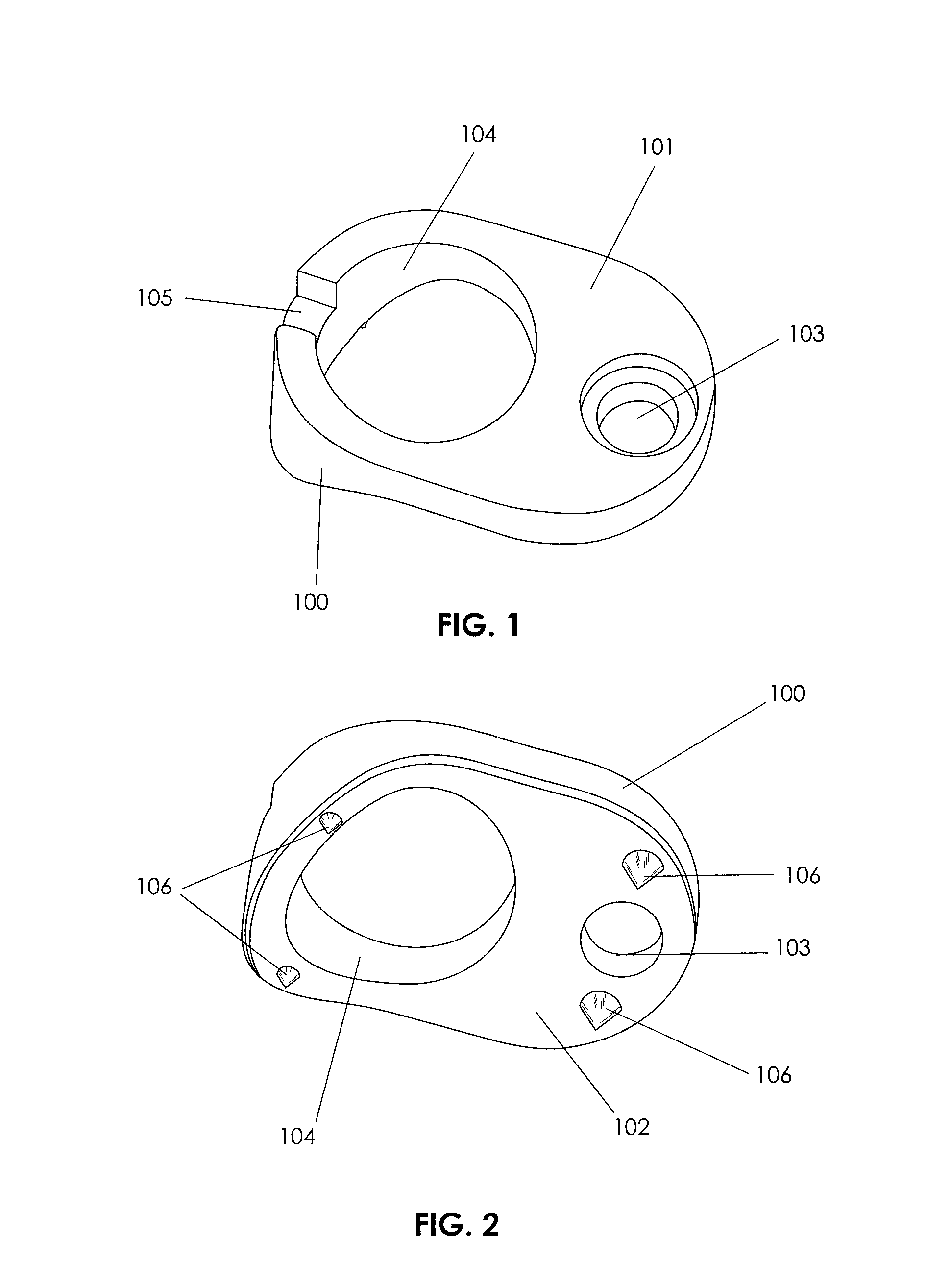
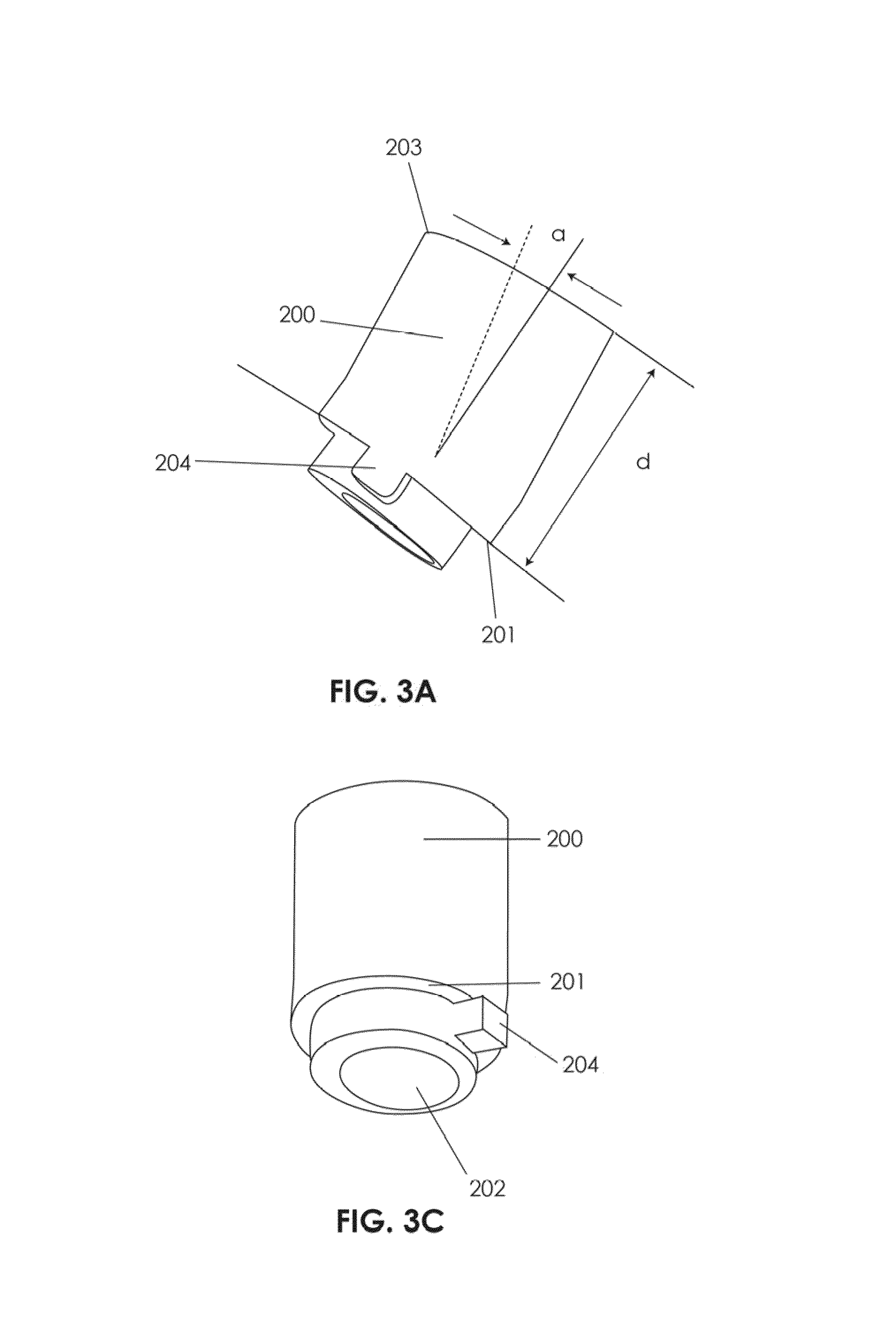
[0089]An inventive surgical system and associated method of use are provided for transcorporeal spinal procedures that create and use an anterior approach to an area in need of surgical intervention, particularly areas at or near a site of neural decompression. Removal or movement of a source of compressing neural pathology is achieved via a surgical access channel created through a single vertebral body instead of through a disc space or through an uncovertebral joint (involving 1 or 2 vertebrae). The access channel has a specifically prescribed trajectory and geometry that places the channel aperture at the posterior aspect of the vertebra in at or immediately adjacent to the targeted compressing pathology, thus allowing the compressing neural pathology to be accessed, and removed or manipulated. The access channel is formed with precise control of its depth and perimeter, and with dimensions and a surface contouring adapted to receive surgical instruments and an implanted bone re...


PUM


Abstract

A system and method are provided for making an access channel through a vertebral body to access a site of neural compression, decompressing it, and repairing the channel to restore vertebral integrity. System elements include an implantable vertebral plate, a guidance device for orienting bone cutting tools and controlling the path of a cutting tool, a bone cutting tool to make a channel in the vertebral body, a tool for opening or partially-resecting the posterior longitudinal ligament of the spine, a tool for retrieving a herniated disc, an implantable device with osteogenic material to fill the access channel, and a retention device that lockably-engages the bone plate to retain it in position after insertion. System elements may be included in a surgery to decompress an individual nerve root, the spinal cord, or the cauda equina when compressed, for example, by any of a herniated disc, an osteophyte, a thickened ligament arising from degenerative changes within the spine, a hematoma, or a tumor.

Description

CROSS REFERENCE TO RELATED APPLICATIONS[0001]This application claims priority to U.S. Provisional Patent Application No. 60 / 972,192 of Lowry et al., entitled “Transcorporeal spinal decompression and repair system and related method”, as filed on Sep. 13, 2007.FIELD OF INVENTION[0002]The invention relates to devices and methods of spinal surgery. More particularly, the invention provides an implant for use in spinal repair surgery and a method for preparing the vertebral volume to receive the implant.INCORPORATION BY REFERENCE[0003]All publications, patents and patent applications mentioned in this specification are herein incorporated by reference to the same extent as if each individual publication, patent or patent application was specifically and individually indicated to be incorporated by reference.[0004]In particular, U.S. patent application Ser. No. 11 / 855,124 of Lowry et al. (filed on Sep. 13, 2007, entitled “Implantable bone plate system and related method for spinal repair...

Claims


Application Information

Patent Timeline
Patent Type & Authority Patents(United States)
IPC IPC(8): A61B17/80
CPCA61B17/1671A61B17/1757A61B17/686A61B17/70A61B17/1637A61F2002/4649A61B17/7059A61B17/809A61B2019/462A61B2090/062
Inventor LOWRY, DAVIDO'FARRELL, DESMONDTUINSTRA, SCOTTVELDMAN, ROGER
Owner GLOBUS MEDICAL INC
Learn More