
External fixation and foot-supporting device

a technology of external fixation and support device, which is applied in the field of therapeutic orthopedic devices, can solve the problems of long treatment duration, cumbersome external fixation structure, patient's foot and lower leg immobilization, etc., and achieve the effects of reducing complexity, reducing pain, and normalizing gai

Inactive Publication Date: 2012-06-05
QUANTUM MEDICAL CONCEPTS
View PDF31 Cites 39 Cited by
  • Summary
  • Abstract
  • Description
  • Claims
  • Application Information

AI Technical Summary

Benefits of technology

"The present invention provides an external fixation system for external fixation of the anatomical skeleton, particularly for patients with foot and ankle problems. The system improves patient comfort, allows for a more normalized gait, reduces deformation of the foot, and non-plantar mounting of a foot ring. The system is easily adjustable, self-removable, and provides improved sensory feedback for a faster recovery. The system includes a base and a plurality of medial-lateral supports that are selectively and releasably coupled to the external fixation frame. The system provides a more normalized gait pattern, reduces stress on the fixation structures, and promotes healing without the need for additional surgical procedures. Overall, the invention provides a more efficient and effective external fixation system for patients with foot and ankle problems."

Problems solved by technology

One drawback of circular fixation treatments includes a lengthy treatment duration—often in excess of one year—during which the patient's foot and lower leg are immobilized in a cumbersome external fixation structure consisting of varied components including pins, wires, support rods, clamps, and frames.
Another drawback of existing circular fixation treatments and systems is that patients have restricted movement.
This restriction is a result of the quantity and placement of cumbersome components that—due to their size, weight, and anatomic placement—severely restrict and impair motion to a range that is far less than the patient's accustomed normal range.
However, existing circular external fixation systems inadequately address the patient's desire and need to use the constricted limb as close to normal as possible.
Specifically, existing external fixation systems inadequately address the patient's need for an ambulatory load-bearing construct that enables or approximates the patient's normal (unencumbered) gait.
Although these solutions provide a “contact” platform under the foot, they fail to provide adequate cushioning, adjustability [for gait], and removability, or any combination of these characteristics.
And, more problematic, injuries and other abnormities of the foot require additional frame elements including a foot ring.
Foot rings can be difficult to align and mount either because of more challenging deformities of the foot such as equinus contractures or varus deformities, or simply because of the difficulty surgeons encounter when trying to manually align the foot ring properly with the foot.
Often the result is an attached foot ring that is non-plantigrade, or poorly aligned with the horizontal axis of the foot.
As a result, the patient often cannot load-bear due to hardware attachments beneath the foot ring, the position of the foot ring, the position of the foot ring relative to the foot, or because of a pre-existing anatomic position that prohibits them from walking normally.
Additionally, many prior-art foot rings, or foot-ring walking attachments, do not provide any cushioning or traction-enhancing features.
Known prior-art or state-of-the-art external fixations systems present additional drawbacks when applied for use as a walking attachment.
Frame members that were not designed specifically as a footplate do not provide for the cushion or tread necessary for such a longitudinal course of treatment.
Moreover, these devices are difficult to adjust to a patient's comfort given the planar variances of each patient's foot position and the need for the device to adapt to a wide spectrum of variation between individual patients.
Further, because the adjustments made to existing devices require tools (such as wrenches), to tighten and loosen nuts on threaded rods and on bolts, it can be very difficult, if not impossible for a patient to accurately adjust their ring for comfort or hygiene.
Any comfort gain by removal for bathing or at bedtime is eclipsed by the arduousness of this task and, often for the entire duration of treatment, an individual patient will not remove their load-bearing ring.
This additional length causes an abnormal gait and further causes hip and knee problems.
The deficiency of this device is multifold in that it nearly eliminates all available attachment points for wire fixation to the foot ring due to it's size and shape that mirrors the foot ring and, hence eclipses, all attachment point options.
The DePuy device, because it attaches using multiple bolts and nuts, makes very difficult if not impossible for the patient to remove his device when bathing or sleeping.
Finally, and most significantly, the DePuy device does not inherently provide a means for planar adjustability; there is no available means for either a surgeon or patient to easily adjust the position of their means for comfort, supplemental correction, or just ambulating.
Problems common to the current-state-of-the-art devices, represented above, include an unacceptable level of precision that must be maintained during the creation of the frame structure in orientating each component while a multitude of fasteners are tightened.
Not only is such adjustments time-consuming, they are often impossible for the patient to make on their own because the patient is unable to reach the fasteners due to poor flexibility or simply because the location is out of the range of normal-human motion for a device worn on the foot.
Moreover, the inter-dependent nature of the fasteners often require incremental adjustments made in sequence with each of the multiple fasteners, which requires a skill beyond the average patient.
Despite the varied attempts at improving external frame fixation systems, many problems specific to the lower extremity and, particularly, to the foot, have not been adequately addressed.
For example, patient comfort, gait, hygiene, ease of removal are inferior in known systems.

Method used


Image

  • External fixation and foot-supporting device
  • External fixation and foot-supporting device
  • External fixation and foot-supporting device

Examples


Embodiment Construction

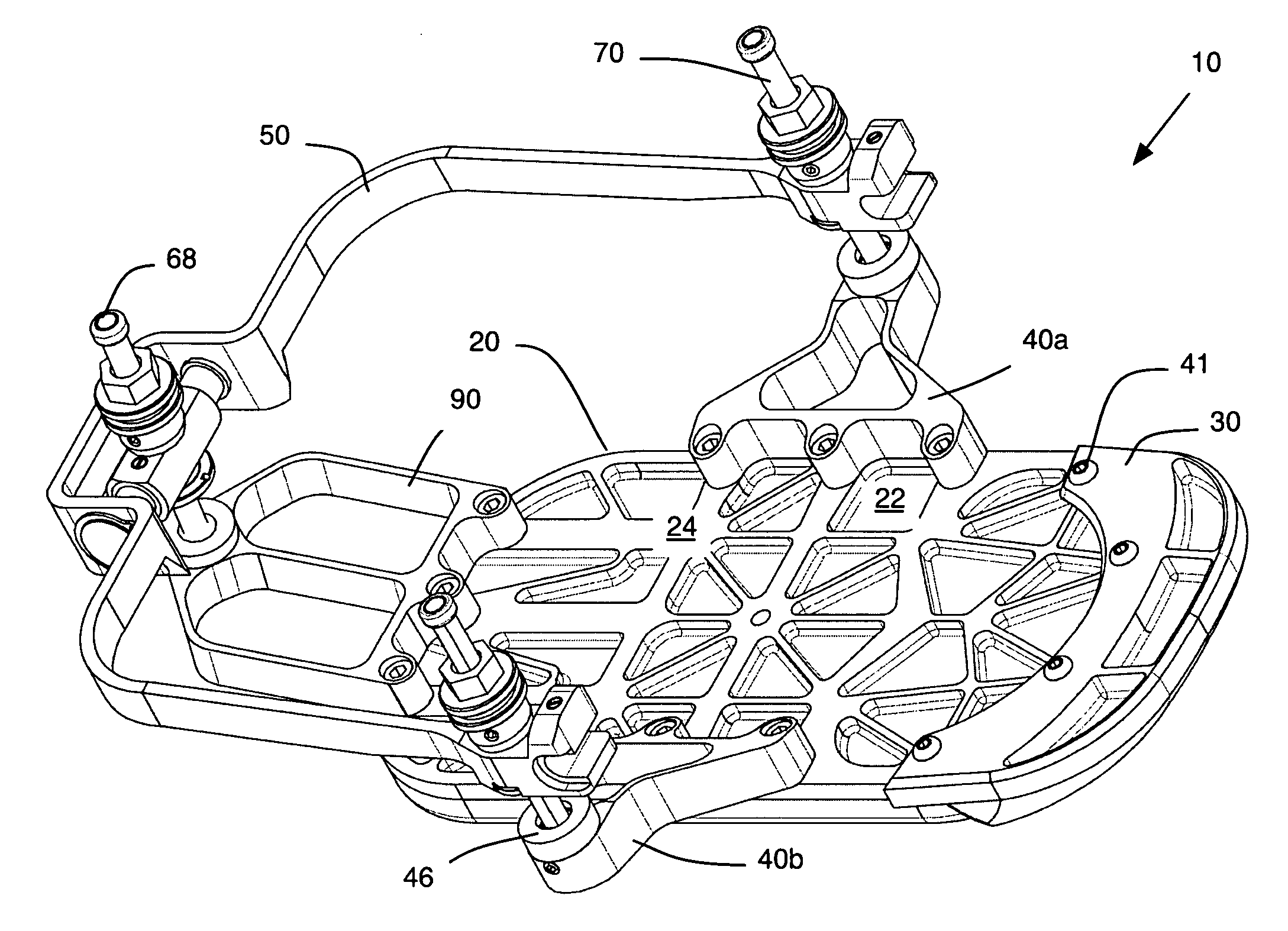
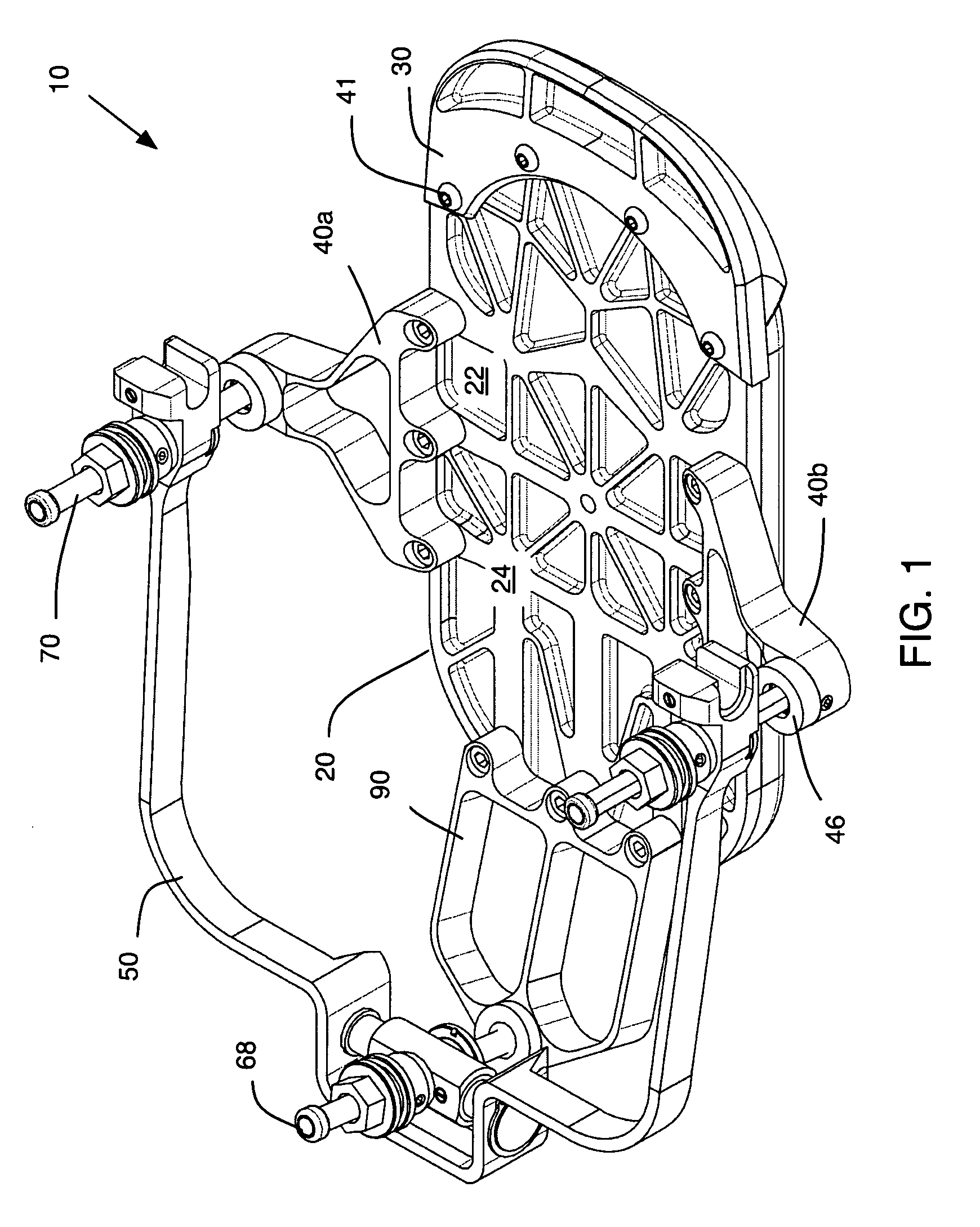
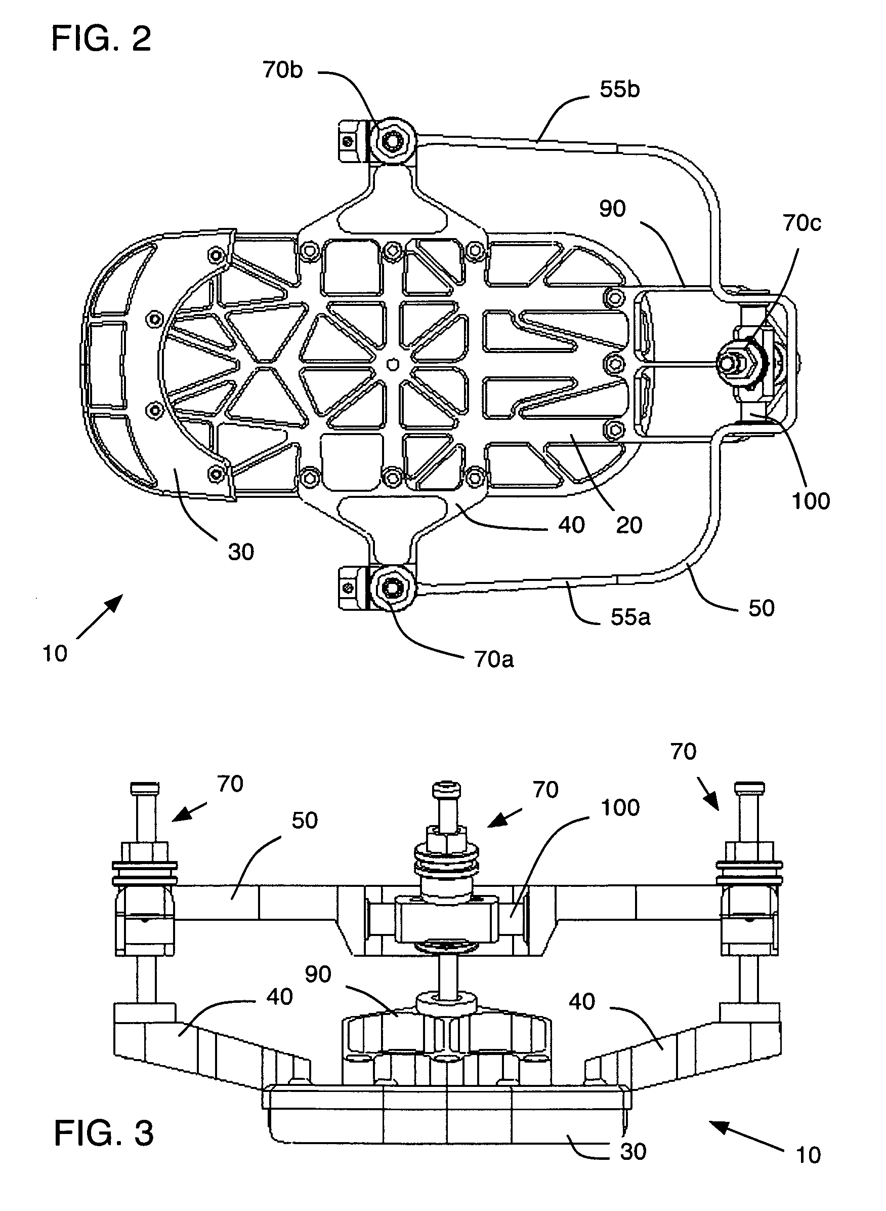
[0077]Possible embodiments will now be described with reference to the drawings and those skilled in the art will understand that alternative configurations and combinations of components may be substituted without subtracting from the invention. Also, in some figures certain components are omitted to more clearly illustrate the invention. In the prior-art, the term “foot-ring” and “foot-plate” often are used synonymously. However, in the present invention an attempt is made to distinguish the known prior art device that surrounds the foot (foot ring) from the present invention that includes components that couple to this foot ring and components adapted for use by the plantar surface of the foot. The later type components are termed foot-plates, or as used herein, a base plate.

[0078]FIGS. 1-4 illustrate a first preferred embodiment of the present invention and provide an overview contextualizing the various components. In this embodiment, a FRAMEWALKER™ brand (available from Quantu...


PUM


Abstract

In one preferred embodiment according to the present invention, an external fixation device for the foot and ankle of the human anatomical skeleton includes a yoke, three adjusters, and a base (or foot plate). The foot plate selectively couples to the yoke without the need of any tools, thereby enabling the patient to self attach and remove the foot plate from an existing circular fixation structure.

Description

PRIORITY CLAIM[0001]The present application claims benefit under 35 USC Section 119(e) of U.S. Provisional Patent Application Ser. No. 61 / 049,865 filed on 2 May 2009. The present application is based on and claims priority from this application, the disclosure of which is hereby expressly incorporated herein by reference.BACKGROUND[0002]The present invention relates to therapeutic orthopedic devices for fixating, immobilizing, and manipulating the human anatomical skeleton, particularly the foot and lower leg. And, more specifically, the present invention relates to external frame systems and devices for fixating lower leg bones and the foot with respect to the tibia.[0003]External Fixation devices locate segments of bone relative to a reference location of the skeleton for varied purposes including reconstruction of fractured or deformed extremities. By distracting or compressing portions of the anatomical skeleton, external fixation devices can correct angulation, rotation, and tr...

Claims


Application Information

Patent Timeline
Patent Type & Authority Patents(United States)
IPC IPC(8): A61B17/00
CPCA61B17/60A61F5/048A61B17/6425A61B17/6491A61F5/0102
Inventor HUEBNER, RANDALL J.SHAEVITZ, ALEXANDER RANDALLMCGRATH, WILLIAMBEAMAN, DOUGLAS N.
Owner QUANTUM MEDICAL CONCEPTS
Learn More