
Method and apparatus for airway detection and segmentation using 3D morphological operators

a morphological operator and airway technology, applied in the field of airway segmentation, can solve the problems of virtually impossible to use a simple threshold to identify all airway voxels within an image, thin or stenosed airways can appear broken or discontinuous, and airway tree segmentation is traditionally a challenging problem

Inactive Publication Date: 2010-08-31
SIEMENS MEDICAL SOLUTIONS USA INC
View PDF2 Cites 7 Cited by
  • Summary
  • Abstract
  • Description
  • Claims
  • Application Information

AI Technical Summary

Benefits of technology

This approach enhances airway segmentation by reducing false positives, maintaining finer details, and reducing computational burden by iteratively growing the initial segmentation with decreasing operator sizes, thus improving the accuracy and efficiency of airway detection.

Problems solved by technology

Airway tree segmentation is traditionally a challenging problem.
While airway voxels have a density near −1000 Hounsfield units (HU), noise and partial volume effects make it virtually impossible to use a simple threshold to identify all airway voxels within an image.
Moreover, due to the size of the voxel, thin or stenosed airways can appear broken or discontinuous.
Finally, image reconstruction artifacts may cause the airways to appear discontinuous.
Such discontinuities may cause problems during the segmentation, potentially resulting in both under- and oversegmentation errors.
Although 3-D region growing is typically extremely fast, it suffers from partial volume effects and noise due to the global threshold used during segmentation.
Further, these methods conventionally lose details, depict incomplete structures, or suffer from parenchymal leakage (“explosion”) to varying degrees.
The disadvantage of this method is the critical dependence on the central axis analysis results, which may be imperfect or fail.
One potential pitfall with this approach is oversegmentation of the airways via false candidates.
Further, most morphology-based methods require heavy computation times.

Method used


Image

  • Method and apparatus for airway detection and segmentation using 3D morphological operators
  • Method and apparatus for airway detection and segmentation using 3D morphological operators
  • Method and apparatus for airway detection and segmentation using 3D morphological operators

Examples


Embodiment Construction

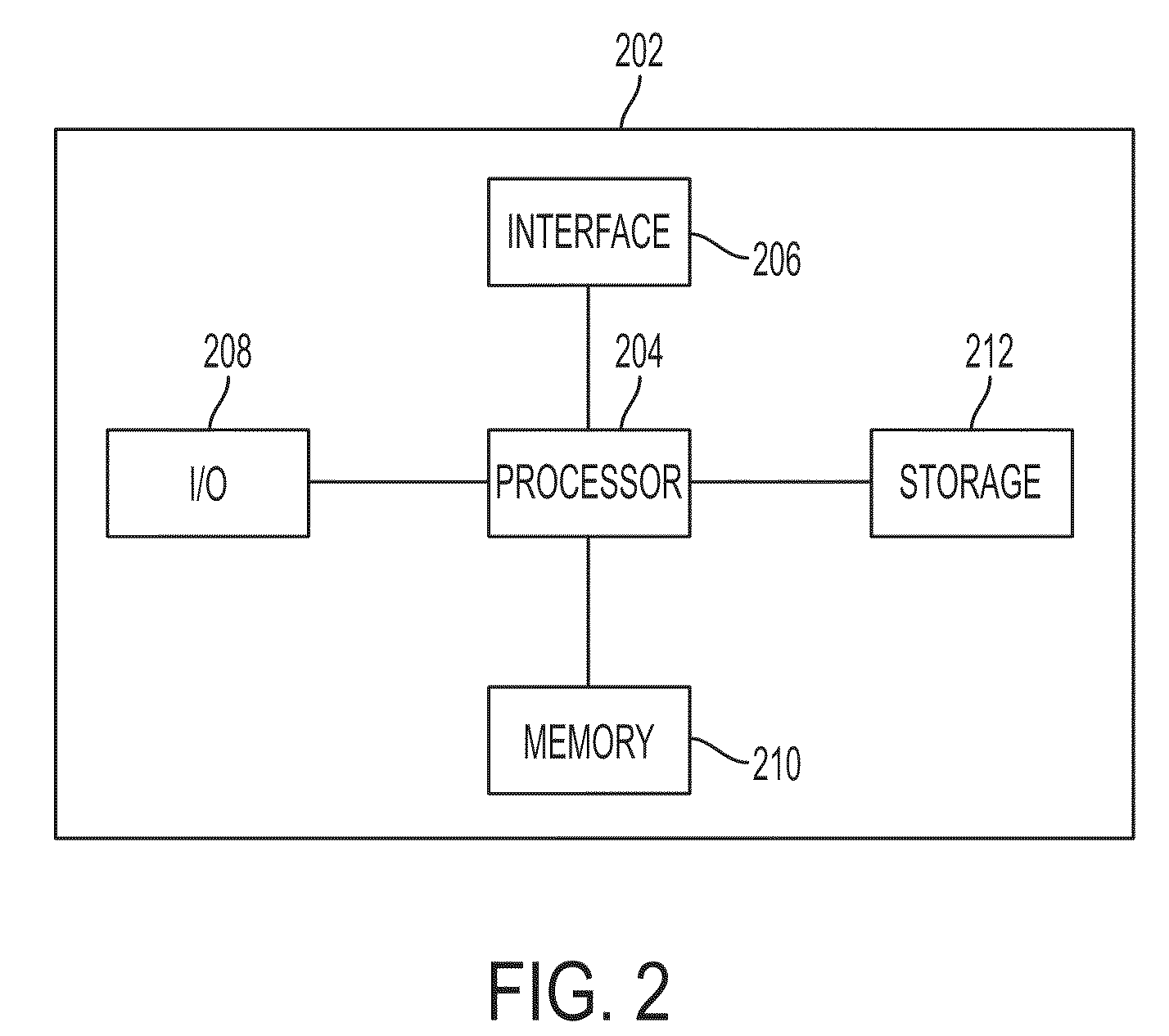
[0031]The following description describes the present invention in terms of the processing steps required to implement an embodiment of the invention. These steps may be performed by an appropriately programmed computer, the configuration of which is well known in the art. An appropriate computer may be implemented, for example, using well known computer processors, memory units, storage devices, computer software, and other components. A high level block diagram of such a computer is shown in FIG. 2. Computer 202 contains a processor 204 which controls the overall operation of computer 202 by executing computer program instructions which define such operation. The computer program instructions may be stored in a storage device 212 (e.g., magnetic disk) and loaded into memory 210 when execution of the computer program instructions is desired. Computer 202 also includes one or more interfaces 206 for communicating with other devices (e.g., locally or via a network). Computer 202 also...


PUM


Abstract

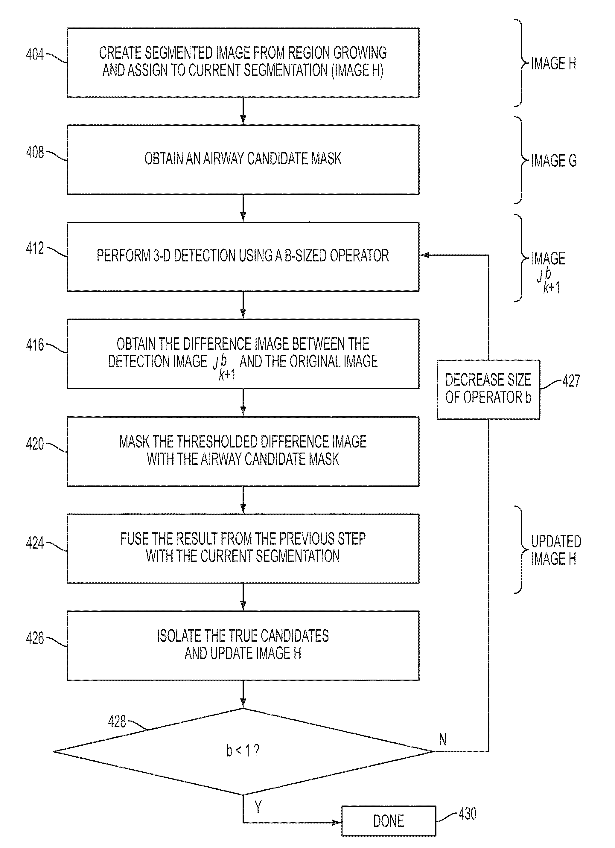
Disclosed is a method and system for detecting a structure (e.g., an airway) within an image. The method and system generate a segmented image from the original (reconstructed) image. The method and system then iteratively apply three dimensional morphological operators of varying sizes to the image to obtain a resulting image. The method and system obtain a candidate mask, apply the candidate mask to the resulting image to generate a masked resulting image, and update the segmented image using the masked resulting image to detect the structure. In one embodiment, the segmented image is generated from region growing. Also disclosed is a method to determine different regions of interest within a given segmented image based on computed projections and boundary lines of the segmented image.

Description

[0001]This application claims the benefit of U.S. Provisional Application No. 60 / 637,805 filed Dec. 21, 2004, which is incorporated herein by reference.BACKGROUND OF THE INVENTION[0002]The present invention relates generally to airway segmentation, and more particularly to using 3-dimensional (3-D) morphology operators for airway detection and segmentation.[0003]A Computed Tomography (CT) scan uses x-ray equipment to obtain image data from different angles around the human body and then uses computer processing of the information to produce cross-sectional images of the body tissues and organs. The image can then be analyzed by methods using morphological operators to highlight specific areas so that radiologists (or other medical personnel) can more easily diagnose problems associated with the patient such as cancers, cardiovascular disease, infectious disease, trauma and musculoskeletal disorders. Current high-resolution CT offers high resolution images of the chest and airways.[0...

Claims


Application Information

Patent Timeline
Patent Type & Authority Patents(United States)
IPC IPC(8): G06K9/00G06V10/34
CPCG06K9/44G06T7/0081G06T7/0091G06K2209/05G06T2207/30101G06T2207/10081G06T2207/30061G06T7/11G06T7/155G06V10/34G06V2201/03
Inventor VAZ, MICHAELKIRALY, ATILLA PETER
Owner SIEMENS MEDICAL SOLUTIONS USA INC
Learn More