X-ray computed tomography apparatus and tomographic method

a computed tomography and computed tomography technology, applied in tomography, applications, instruments, etc., can solve the problems of insufficient x-ray intensity distribution data required for reconstructing a heart tomographic image, image cannot be reconstructed in the expected temporal resolution, and scan schedule cannot be changed

Active Publication Date: 2010-07-13
TOSHIBA MEDICAL SYST CORP
View PDF15 Cites 4 Cited by
  • Summary
  • Abstract
  • Description
  • Claims
  • Application Information

AI Technical Summary

Benefits of technology

[0031]One of the main features according to the present invention is that collection of data required for electrocardiogram-gated reconstruction is ensured, and also an X-ray dosage can be reduced. To explain it simply, the X-ray CT apparatus according to the first embodiment detects a predetermined characteristic wave from each electrocardiographic waveform acquired from the electrocardiograph. For example, according to the embodiment, the X-ray CT apparatus detects an R-wave as a characteristic wave in an electrocardiographic waveform, and acquires the time of the detection (for example, a time distance from the start of acquisition of the electrocardiographic waveform to the detection).

Problems solved by technology

If a heartbeat abnormality, such as arrhythmia caused by extrasystole or a heartbeat fluctuation, occurs, there is a possibility that sufficient X-ray intensity distribution data required for reconstructing a heart tomographic image are not obtained by scanning the heart even in accordance with the scanning schedule.
However, as shown in FIG. 9B, when an interval between R-waves changes substantially during X-ray irradiation due to an abnormal heartbeat, there is a problem that even if the X-ray irradiation is performed in accordance with the determined scanning schedule, intended X-ray intensity distribution data of two normal heartbeats cannot be obtained, consequently an image cannot be reconstructed in the expected temporal resolution. FIGS. 9A to 9D are schematic diagrams for explaining a conventional technology.
According to the conventional technology, there is a problem that the scanning schedule cannot be changed in response to an abnormal heartbeat occurring before the scanning, because data acquired from the electrocardiograph is not monitored after the scanning schedule is determined until actually starting the scanning, as a result, an X-ray dosage tends to be large to acquire a required amount of intended X-ray intensity distribution data in a specific phase.
For example, as shown in FIG. 9D, if an abnormal heartbeat occurs before scanning, an actual electrocardiographic waveform deviates from an expected electrocardiographic waveform in a starting period of X-ray irradiation in the determined scanning schedule, as a result, an X-ray dosage tends to be large to acquire intended X-ray intensity distribution data of two normal heartbeats in a specific phase, which is a problem.
In other words, the conventional technology has a problem that even if an abnormal heartbeat occurs before scanning, a required amount of intended X-ray intensity distribution data in a specific phase can be acquired; however, the scanning schedule cannot be changed in response to the abnormal heartbeat occurring before the scanning, thereby causing an unnecessary exposure to X-rays.
As described above, the Retrospective Gating method has the problem that an X-ray dosage tends to be large to acquire a required amount of intended X-ray intensity distribution data in a specific phase, and also the Prospective Gating method has a similar problem.

Method used


Image

  • X-ray computed tomography apparatus and tomographic method
  • X-ray computed tomography apparatus and tomographic method
  • X-ray computed tomography apparatus and tomographic method

Examples


first embodiment

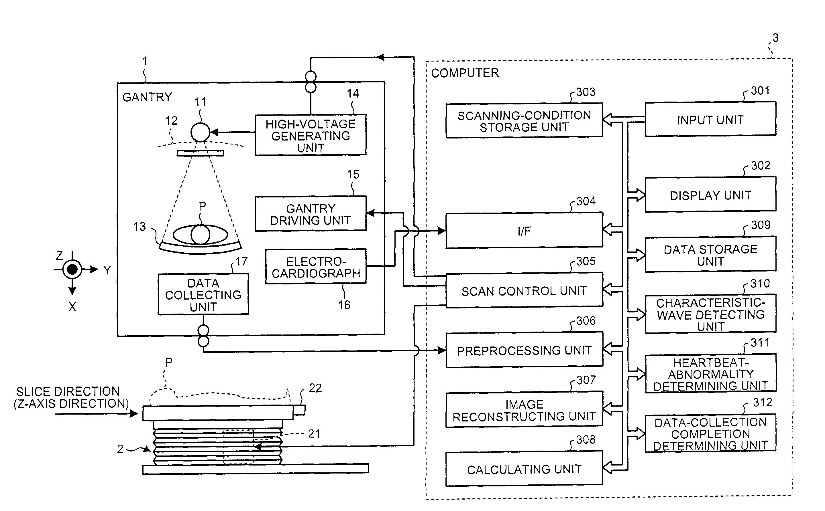
Outline and Features of X-ray CT Apparatus

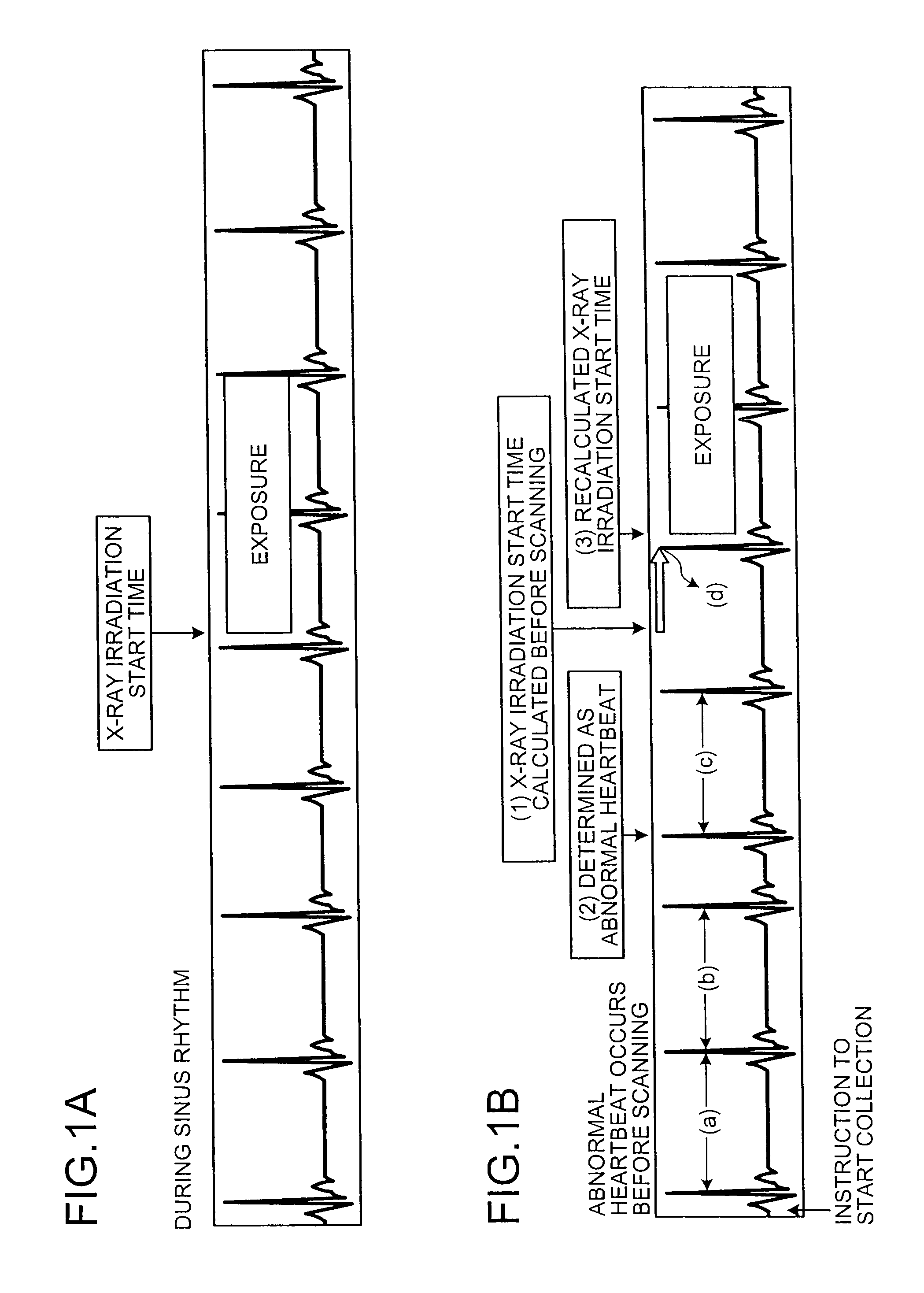
[0027]First of all, main features of an X-ray CT apparatus according to a first embodiment of the present invention are specifically explained below with reference to FIGS. 1A, 1B and 2. FIGS. 1A and 1B are schematic diagrams for explaining an outline and feature of the X-ray CT apparatus according to the first embodiment, and FIG. 2 is a schematic diagram for explaining features of the X-ray CT apparatus according to the first embodiment.

[0028]The X-ray CT apparatus according to the first embodiment is configured to start tomographic scanning that includes irradiating with X-rays a subject attached with an electrocardiograph, detecting X-rays passed through the subject, and reconstructing a heart tomographic image of the subject based on X-ray intensity distribution data that is information about the detected X-rays, in accordance with a scanning start time obtained from an electrocardiographic waveform acquired from the electrocardiograph....

second embodiment

Outline and Features of X-Ray CT Apparatus

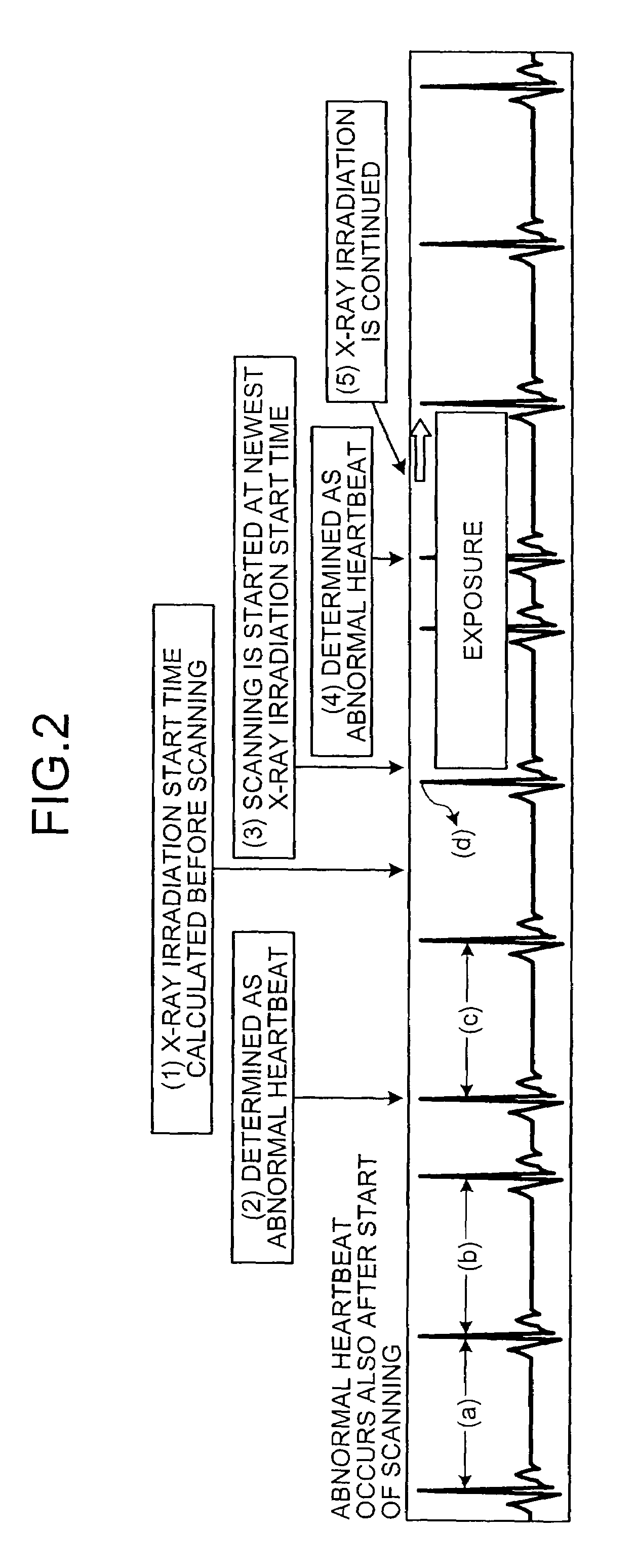
[0096]First of all, main features of an X-ray CT apparatus according to a second embodiment of the present invention are specifically explained below with reference to FIGS. 5A, 5B, 6A and 6B. FIGS. 5A and 5B are schematic diagrams for explaining an outline of the X-ray CT apparatus according to the second embodiment, and FIGS. 6A and 6B are schematic diagrams for explaining features of the X-ray CT apparatus according to the second embodiment.

[0097]The X-ray CT apparatus according to the second embodiment is configured to collect X-ray intensity distribution data within a certain collection period obtained from an electrocardiographic waveform acquired from an electrocardiograph, during tomographic scanning that includes irradiating with X-rays a subject attached with the electrocardiograph, detecting X-rays passed through the subject, and reconstructing a heart tomographic image of the subject based on detected X-ray intensity distribution...


PUM


Abstract

An X-ray CT apparatus detects a predetermined characteristic wave from each electrocardiographic waveform acquired from an electrocardiograph, predicts intervals with which the characteristic wave appears based on appearance times of the detected predetermined characteristic wave in each electrocardiographic waveform, and determines an X-ray irradiation start time based on the predicted interval time. After an instruction to start collection of X-ray intensity distribution data is received, if an appearance interval of a detected predetermined characteristic wave is within an acceptable normal-heartbeat range, it is determined as a normal heartbeat. If the appearance interval of the detected predetermined characteristic wave is outside the acceptable normal-heartbeat range, it is determined as an abnormal heartbeat. During a period after receiving the instruction to start the collection until reaching a time of starting the collection of X-ray detection information, when determined as an abnormal heartbeat, an X-ray irradiation start time is recalculated, and then determined.

Description

CROSS-REFERENCE TO RELATED APPLICATIONS[0001]This application is based upon and claims the benefit of priority from the prior Japanese Patent Application No. 2007-223035, filed on Aug. 29, 2007; the entire contents of which are incorporated herein by reference.BACKGROUND OF THE INVENTION[0002]1. Field of the Invention[0003]The present invention relates to an X-ray computed tomography apparatus and a tomographic method.[0004]2. Description of the Related Art[0005]Conventionally, an X-ray Computed Tomography apparatus (hereinafter, “X-ray CT apparatus”) plays an important role in various medical practices, such as a diagnosis of a disease, a treatment, and an operation plan, by reconstructing and providing a tomographic image that indicates morphologic information about human body tissue in a subject body on a plane (fault plane) irradiated with an X-ray based on X-ray intensity distribution data that is obtained by detecting the intensity of an X-ray passed through the subject.[0006]...

Claims


Application Information

Patent Timeline
Patent Type & Authority Patents(United States)
IPC IPC(8): A61B6/03H05G1/60
CPCA61B5/0456A61B6/503A61B6/032A61B5/352
Inventor MUKUMOTO, GOKAMINAGA, SHIGEOYASUDA, HISASHIYAMAHANA, MASAOMORINO, KATSUHIROSETO, HIROMITSU
Owner TOSHIBA MEDICAL SYST CORP
Why Eureka
Try Eureka