
Marking catheter for placement using frameless stereotaxy and use thereof

a technology of frameless stereotaxy and positioning catheter, which is applied in the field of surgical devices and appliances, can solve the problems of cumbersome stereotactic frame in general, cumbersome stereotactic frame in particular, and reduce the likelihood of healthy surrounding brain tissue being damaged, and achieve the effect of minimizing the likelihood of damag

Inactive Publication Date: 2007-12-25
WASCHER THOMAS M
View PDF5 Cites 21 Cited by
  • Summary
  • Abstract
  • Description
  • Claims
  • Application Information

AI Technical Summary

Benefits of technology

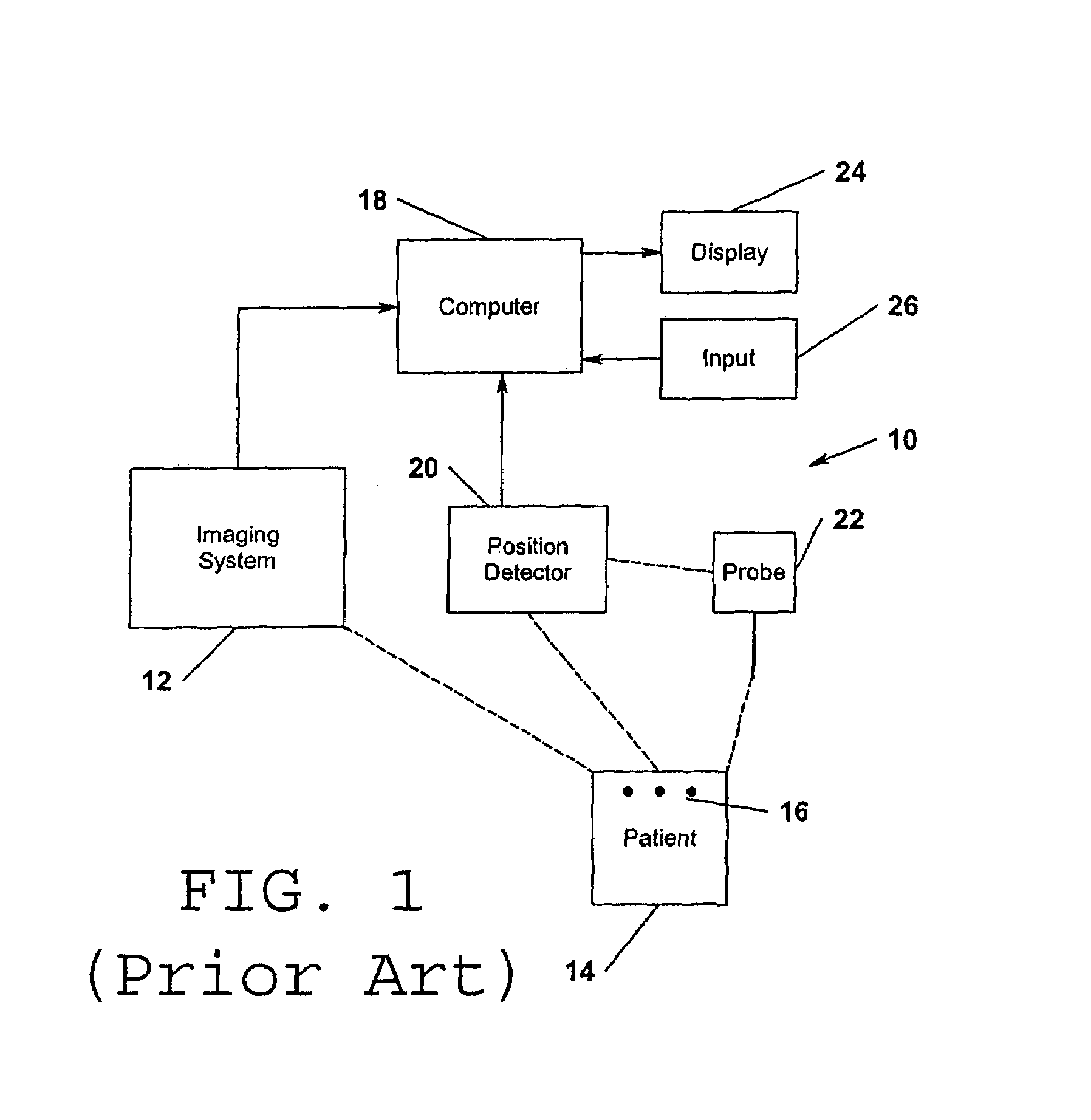
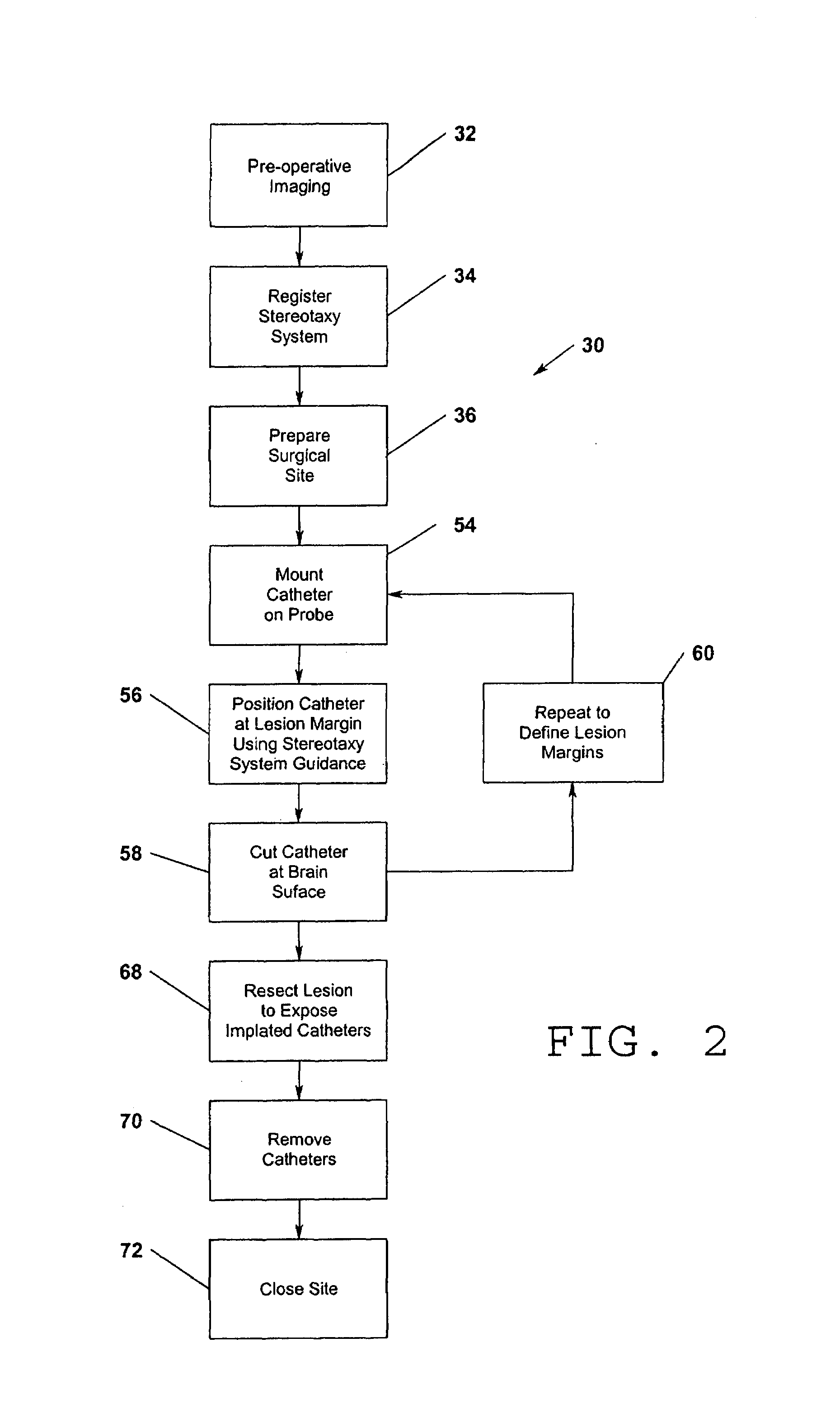
[0014]The present invention allows a neurosurgeon more accurately and completely to remove a brain lesion while minimizing the likelihood that healthy surrounding brain tissue will be damaged by the procedure. The present invention allows a neurosurgeon to take full advantage of pre-operative brain imaging, obtained using computed tomography (CT) or magnetic resonance imaging (MRI) scans, in combination with frameless stereotaxy accurately to define the boundaries of the lesion such that the lesion accurately can be resected during the surgical procedure. In accordance with the present invention, the lesion margins are defined in a manner such that brain-shift that may occur during the resection does not affect the ability of the surgeon accurately and completely to remove the tumor. In accordance with the present invention, one or more thin and flexible catheters are placed into the brain, using a frameless stereotaxy system for guidance before the resection has begun, to define the margins of the lesion to be resected. The catheters are designed to fit over a frameless stereotaxy system localizing probe and, using a frameless stereotactic localization procedure, are placed in positions to define the margins of the lesion in question. The resection then proceeds as normal, e.g., using ultrasonic resection, until the catheters are exposed at all margins of the resection cavity. In this fashion the surgeon is assured that all of the lesion is resected. The catheters will move with the surrounding brain as the brain shifts during the course of the resection, negating the affect of intra-operative brain-shift. The localizing catheters are removed after the resection is completed.
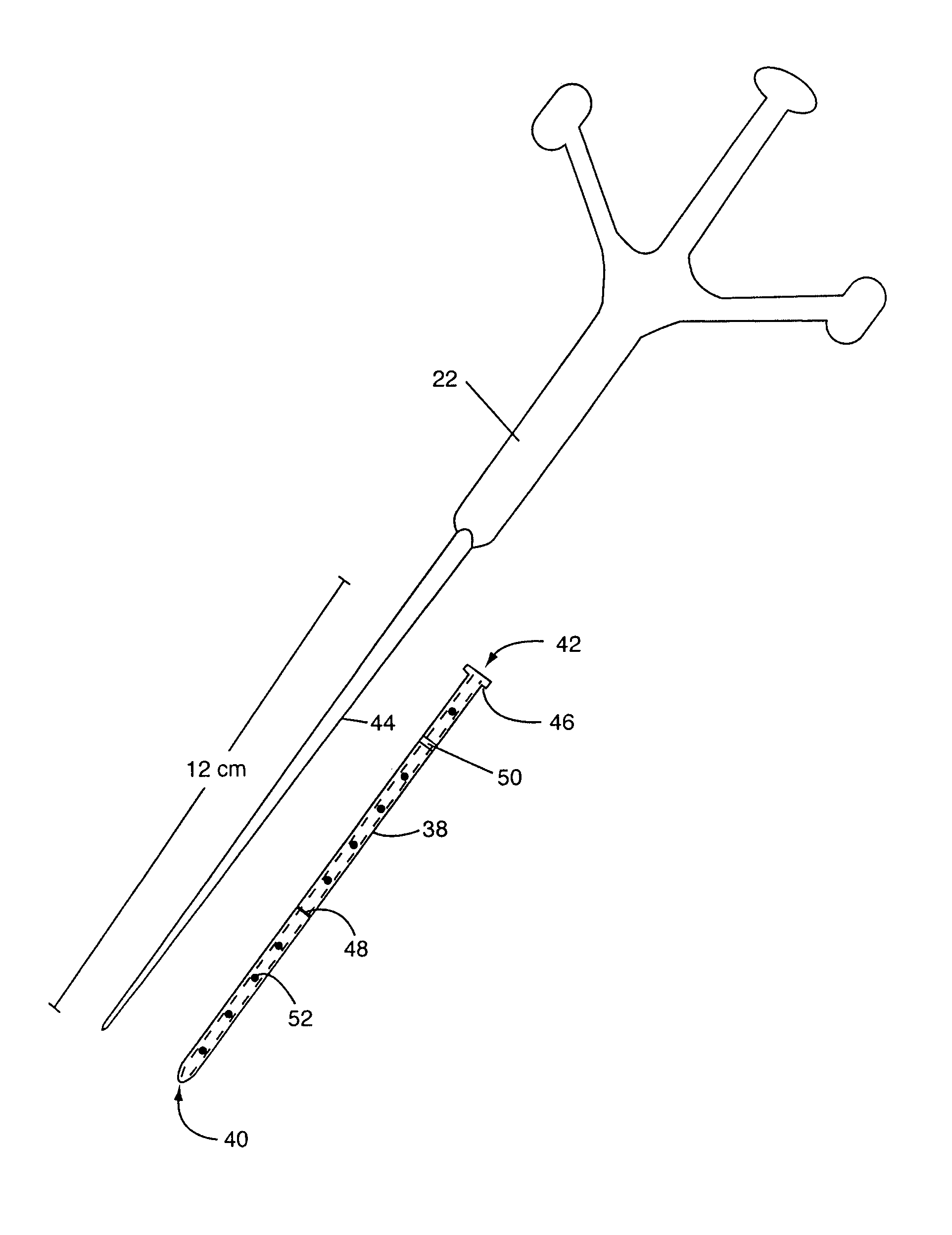
[0017]After preparing the surgical site, e.g., to expose the area of the brain upon which surgery is to take place, the surgeon mounts a marking catheter in accordance with the present invention onto the frameless stereotaxy system probe. In accordance with the present invention, the marking catheter preferably is made of a thin soft or flexible material, such as silicone rubber (e.g., Silastic). The marking catheter is sized to fit over the frameless stereotaxy system localizing probe with a closed end of the catheter at the distal end of the probe. A flange may be provided at the opposite open end of the catheter to aid in manually sliding the catheter off the probe once it is in position. The external surface of the catheter preferably may be marked with length indicators, e.g., rings and / or dots, to assist the surgeon in visualizing the depth of the target lesion. The catheter preferably may be made of a brightly colored material to enhance visualization thereof during the operative procedure.
[0019]Having positioned one or more marking catheters in accordance with the present invention into the brain to define the margins of the lesion to be resected, the resection of the lesion may proceed in a normal manner, e.g., using conventional micro-surgical techniques. Tumor removal continues until the marking catheters that define the margins of the lesion have been exposed down to their tips. Since the marking catheters are soft and pliable, they will move as the surrounding brain shifts into the cavity of tumor resection, maintaining their positions at the lesion margins, thereby virtually eliminating the error associated with brain-shift. Once all of the catheters have been exposed by way of tumor resection, the surgeon is assured that gross total tumor removal has been accomplished with minimal damage to surrounding normal tissue. At this point the marking catheters can be removed, and the procedure completed in the normal manner.

Problems solved by technology

Stereotactic systems using stereotactic frames have a series of limitations.
Stereotactic frames are cumbersome in general.
They are especially cumbersome for procedures requiring more than a few target points and in volumetric stereotactic procedures where the demands of the procedure dictate the need for a larger working area, yet where such demands come into conflict with the physical structure of the frame.
A conventional stereotactic frame is typically a cage structure extending about the patient's head that inherently restricts freedom of movement of the surgeon.
Many such mechanical adjustments become very cumbersome when a surgeon is confronted with an infinite number of points from which to define a boundary of a volumetric lesion.
Some surgeons find the stereotactic frame application procedure difficult and time consuming.
Patients also find this uncomfortable.
In addition, the necessity to repeat CT and MRI examinations with the frame attached for obtaining pre-operative images increases the cost to patients.
Brain-shift is a major limitation of all neuro-navigational systems that rely on pre-operative imaging data.
With ongoing surgery a remarkable deformation of the brain tissue may occur, resulting in navigational localizing errors as the real position of brain structures varies from that indicated in the pre-operative imaging.
Shifts of a deep tumor margin can lead to incomplete or too-deep resection.
As tumor resection proceeds, brain-shift may result in significant inaccuracy of the navigation system for a high percentage of patients, so that as the operation proceeds some correction of the navigation system must be performed if it is to be relied upon.
It is not only cost intensive but also time consuming.
However, there are inter-individual differences in brain-shift behavior, and brain tissue properties might change during surgical procedures, because of changes in intra-cranial pressure or blood carbon-dioxide concentrations or because of alterations of the water content of the brain after the administration of osmotically active drugs.

Method used


Image

  • Marking catheter for placement using frameless stereotaxy and use thereof
  • Marking catheter for placement using frameless stereotaxy and use thereof
  • Marking catheter for placement using frameless stereotaxy and use thereof

Examples


Embodiment Construction

[0027]The present invention provides for the improved use of frameless stereotaxy systems to improve surgical results. The present invention will be described in detail herein with reference to use thereof for the accurate and complete resection of brain lesions. In this application, the present invention employs a frameless stereotaxy or neuro-navigation system to define accurately the margins of the lesion to be resected in a manner such that the error typically associated with brain-shift occurring during lesion removal is eliminated without requiring expensive and complicated intra-operative imaging. In this way complete tumor removal can be assured with minimized impact on surrounding normal tissue. It should be understood, however, that the present invention may be applicable to use in other surgical contexts, such as the removal of tumors in all areas of the human and / or animal body and / or other surgical procedures on humans and / or animals.

[0028]The present invention is emplo...


PUM


Abstract

A marking catheter made out of a flexible material and having a closed distal end and an open proximal end is provided. The marking catheter is sized and shaped to fit over the probe of a frameless stereotaxy system. One or more marking catheters may be positioned using the frameless stereotaxy system to define accurately an area identified in pre-operative imaging. For example, a plurality of marking catheters may be used to define physically the margins of a brain lesion. The brain lesion may then be removed to expose the positioned catheters, thereby assuring complete and effective lesion removal. The flexible catheters will move with any brain-shift occurring during lesion removal, thereby to maintain an accurate indication of the lesion margin.

Description

FIELD OF THE INVENTION[0001]The present invention pertains generally to surgical devices and appliances and, more particularly, to such devices as used in association with frameless stereotaxy and other systems used for performing surgery on the brain and other parts of the body, and methods for using such devices and appliances.BACKGROUND OF THE INVENTION[0002]Stereotaxy refers to a technique, most often applied to the nervous system, in which the contents of a patient's skull (or other portion of a patient's body) are considered in a precise three-dimensional space defined by a measuring instrument. Traditionally, the measuring instrument used to perform stereotaxy is a stereotactic frame, typically taking the form of a cage structure, that is fixed to the patient's skull or other part of the patient's body. Stereotactic frames are mechanical devices typically based upon a Cartesian or polar coordinate system. These systems typically include a means for securing the stereotactic f...

Claims


Application Information

Patent Timeline
IPC IPC(8): A61B19/00
CPCA61B19/52A61B19/5244A61B19/54A61B2019/5487A61B2019/5255A61B2019/5408A61B2019/462A61B90/36A61B2034/2055A61B2090/062A61B34/20A61B90/39A61B2090/3908A61B2090/3987
Inventor WASCHER, THOMAS M.
Owner WASCHER THOMAS M
Learn More