
Methods for diagnosis of low grade astrocytoma

a low-grade astrocytoma and low-grade astrocytoma technology, applied in the field of low-grade astrocytoma diagnosis, can solve the problems of difficult to distinguish low-grade astrocytoma cells from normal quiescent or reactive cells, difficult to detect low-grade astrocytoma cells, and small abnormal appearance of low-grade astrocytoma cells

Inactive Publication Date: 2007-10-09
THE GOVERNORS OF THE UNIV OF ALBERTA +1
View PDF1 Cites 27 Cited by
  • Summary
  • Abstract
  • Description
  • Claims
  • Application Information

AI Technical Summary

Benefits of technology

This method enables efficient and reliable differentiation of low-grade astrocytoma cells from normal astrocytes, facilitating earlier diagnosis and treatment by using immunohistochemical, immunofluorescence, immunoelectron microscopy, and immunoblotting techniques with anti-J1-31 antibodies, improving patient prognosis.

Problems solved by technology

Morphologically, low-grade astrocytomas are usually normal in appearance, making it difficult to distinguish low-grade astrocytoma cells from normal quiescent or reactive cells.
Some grade 2 low-grade astrocytomas may be slightly abnormal in appearance, but are not easily detected by routine microscopic evaluation of tissue biopsy samples.
These antibodies typically react with both astrocytes and astrocytoma cells, and thus do not serve to differentiate between normal and transformed cells beyond facilitating morphological inspection
Although the prognosis for patients with grade 1 astrocytoma is good, with some patients known to live 30 years or more following diagnosis, the prognosis for patients with grade 2 low-grade astrocytoma is much less optimistic.

Method used


Image

  • Methods for diagnosis of low grade astrocytoma

Examples


example 1

Localization of J1-31 Antigen and Glial Fibrillary Acidic Protein (GFAP) in Formalin-Fixed / Paraffin-Embedded Tissue Sections Using Immunohistochemical Staining

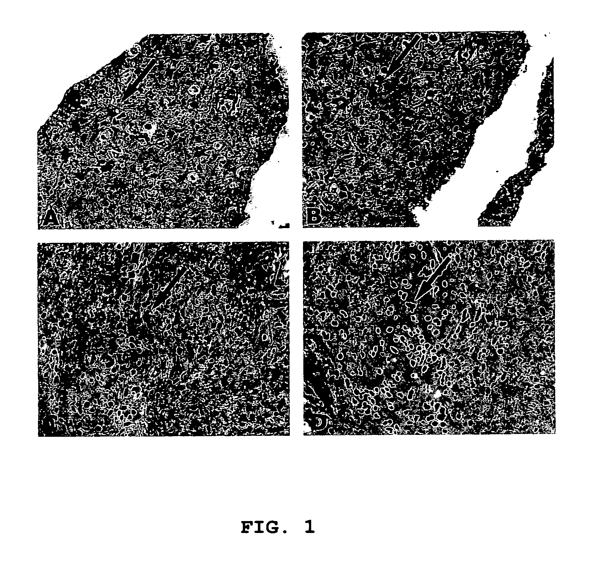
[0076]Formalin-fixed and paraffin-embedded tissue sections of a low grade astrocytoma were prepared and stained, as described above. Briefly, the tissue sections were treated as follows:[0077]1. Slides of the tissue sections were warmed overnight at 60° C.[0078]2. The slides were incubated in Histoclear for 5 minutes; repeated once.[0079]3. The slides were incubated in absolute ethanol for 3 minutes; repeated once.[0080]4. The slides were incubated in 95% ethanol for 3 minutes; repeated once.[0081]5. The slides were incubated in H2O2:methanol for 20 minutes.[0082]6. The slides were washed in DPBS for 5 minutes.[0083]7. The slides were treated in antigen unmasking solution (0.1 M sodium citrate buffer, pH 6.0) in a pressure-cooker and heated in a microwave oven at 600 W twice for 15 minutes each.[0084]8. The slides were left in...


PUM

PropertyMeasurementUnit
pHaaaaaaaaaa
volumeaaaaaaaaaa
volumeaaaaaaaaaa

Abstract

A method for identifying low grade astrocytoma cells in a sample is provided, wherein the method distinguishes between low grade astrocytoma cells and normal astrocytes, thus permitting early diagnosis of astrocytoma. The method uses antibody directed against J1-31 to test astrocytes in the sample for the presence or absence of J1-31 polypeptide, the low grade astrocytoma cells being characterized by the absence of J1-31 while normal astrocytes are characterized by the presence of J1-31.

Description

CROSS-REFERENCE TO RELATED APPLICATION(S)[0001]This application is a divisional application of U.S. patent application Ser. No. 10 / 269,773 filed Oct. 10, 2002, now abandoned which claims the benefit of U.S. Provisional Application Ser. No. 60 / 328,917, filed Oct. 11, 2001, the content of which is hereby incorporated by reference in its entirety.FIELD OF THE INVENTION[0002]The subject of the present invention is a method for using an antibody to detect the presence of a disease, and more specifically to distinguish low grade astrocytomas from normal reactive astrocytes.BACKGROUND OF THE INVENTION[0003]Astrocytomas are members of the glioma family of tumors; that is, tumors arising from glial cells. In the case of astrocytomas, the tumors arise from a type of glial cell called astrocytes.[0004]Astrocytes, named by Spanish neuroanatomist Santiago Ramon y Cajal in 1913 after he discovered the star-shaped cells, are the major cell types of the central nervous system that responds to vario...

Claims


Application Information

Patent Timeline
Patent Type & Authority Patents(United States)
IPC IPC(8): G01N33/53C07K16/30C12Q1/68G01N33/564G01N33/574
CPCG01N33/57407
Inventor IMAM, S. ASHRAFMALHOTRA, SUDARSHAN
Owner THE GOVERNORS OF THE UNIV OF ALBERTA
Learn More