Method of treating a patient with a neurodegenerative disease using ultrasound
a neurodegenerative disease and ultrasound technology, applied in the field of ultrasound treatment of patients with neurodegenerative diseases, can solve the problems of poor tissue penetration, limiting therapeutic success rate, and limiting benefit-to-risk ratio, so as to accelerate wound healing, accelerate healing, and accelerate healing of various types
- Summary
- Abstract
- Description
- Claims
- Application Information
AI Technical Summary
Benefits of technology
Problems solved by technology
Method used
Image
Examples
example 1
Materials and Methods
[0038]Animal Preparation: Rabbits were anesthetized with ketamine (60 mg / kg), xylazine (6 mg / kg) and chlorpromazine (25 mg / kg), and sedation was maintained with sodium pentobarbital as needed. The femoral arteries were dissected 5 cm distal to the origin of the superficial branch, and the profunda femorus and superficial arteries were ligated close to their origin. A Doppler flow probe was placed distally around the isolated segment and two parallel ligatures were placed around the femoral artery 1 cm distal to the profunda branch. These reduced flow by approximately 50% remained in place for the duration of the experiment. Following this, of filter paper saturated with 20% ferric chloride was placed on the femoral artery and thrombosis was assessed by monitoring flow which approached 0 after occlusion. In some animals a completely occlusive suture was placed around the artery.
[0039]Experimental Protocol: Rabbits were assigned to receive: 1) ultrasound alone, 2)...
example 2
Treatment of Surgically Induced Arterial Occlusion with Ultrasound
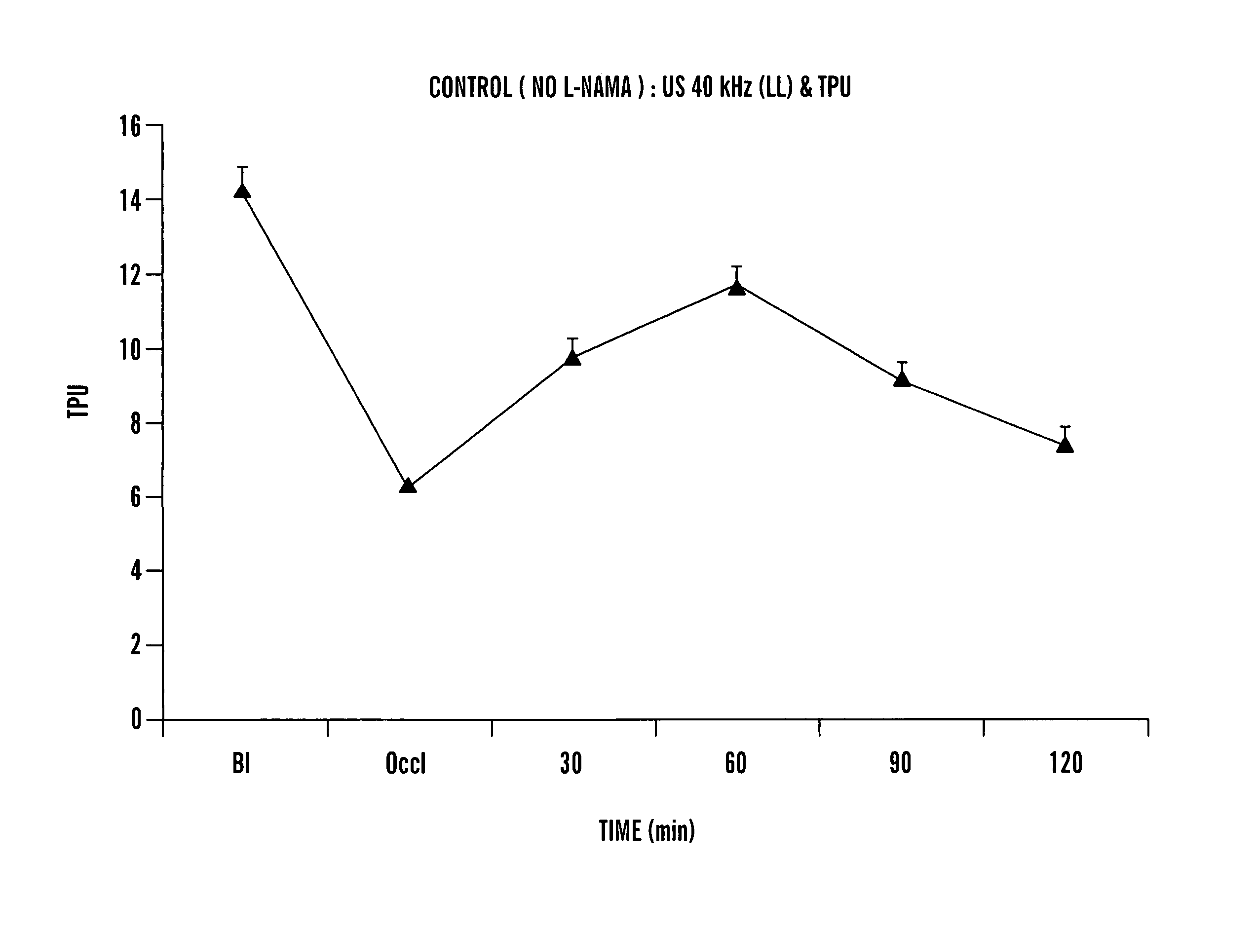
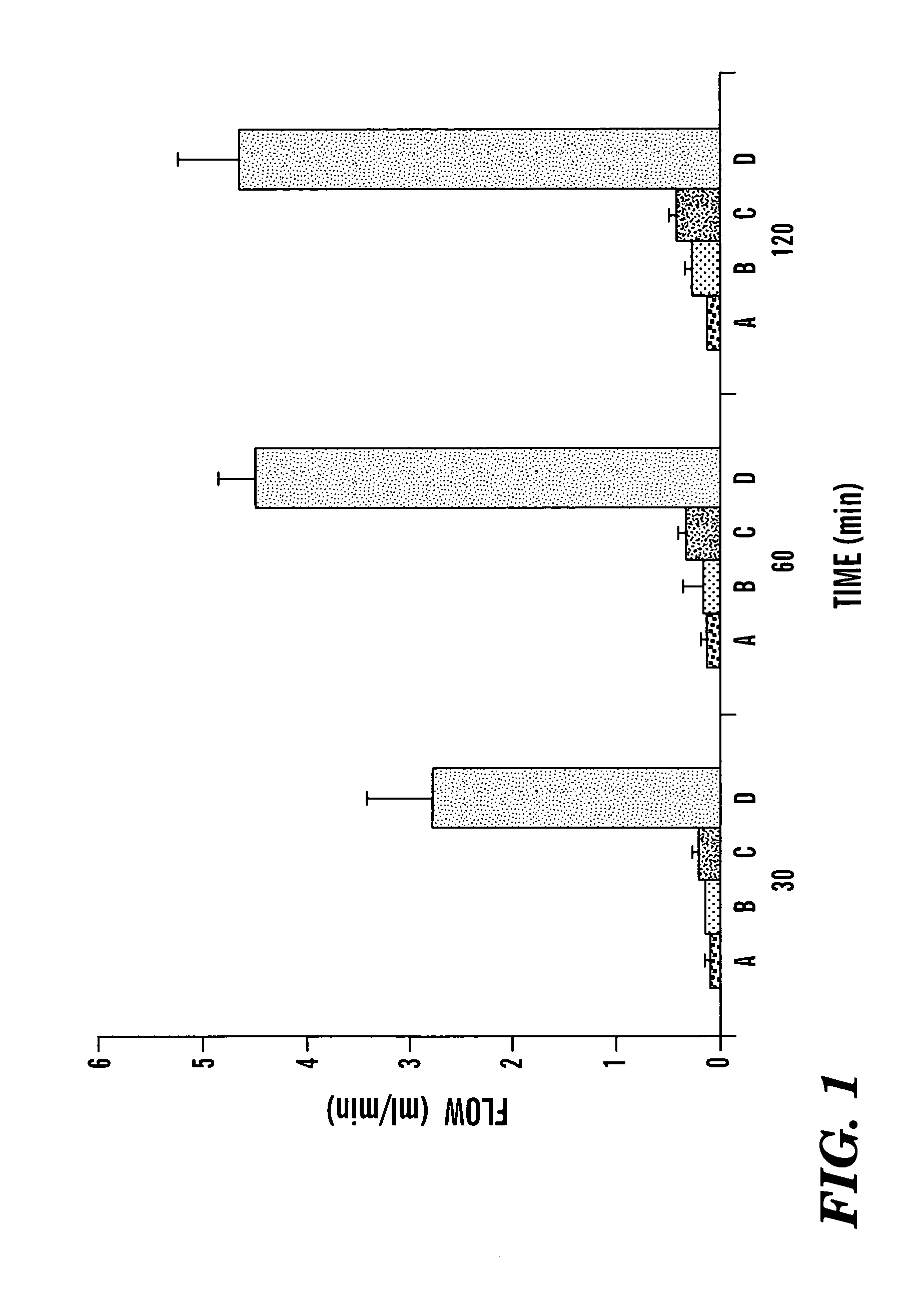
[0042]Occlusive thrombi formed in all femoral arteries within 20–30 min of placement of the constriction and application of 20% ferric chloride. Arterial flow was 12.0±0.7 ml / min at baseline, declined to 5.8±0.4 after placement of the constriction and was 0.1±0.1 after thrombosis. Three different treatment regimens were administered following thrombosis: ultrasound alone, streptokinase alone or the combination of streptokinase and US. Flow in control vessels receiving no treatment remained at near 0 after 30, 60, and 120 min (FIG. 1). Treatment with ultrasound alone resulted in no significant increase in flow, whereas treatment with streptokinase alone resulted in a small but significantly increased flow at 120 min to 0.4±0.1 ml / min (p<0.001). The combination of streptokinase and ultrasound resulted in greater reperfusion, with flow of 2.6±0.7 ml / min at 30 min, 4.6±0.4 ml / min at 60 min and 4.8±0.6 ml / min at 120 min. T...
example 3
Monitoring of Heating by Ultrasound
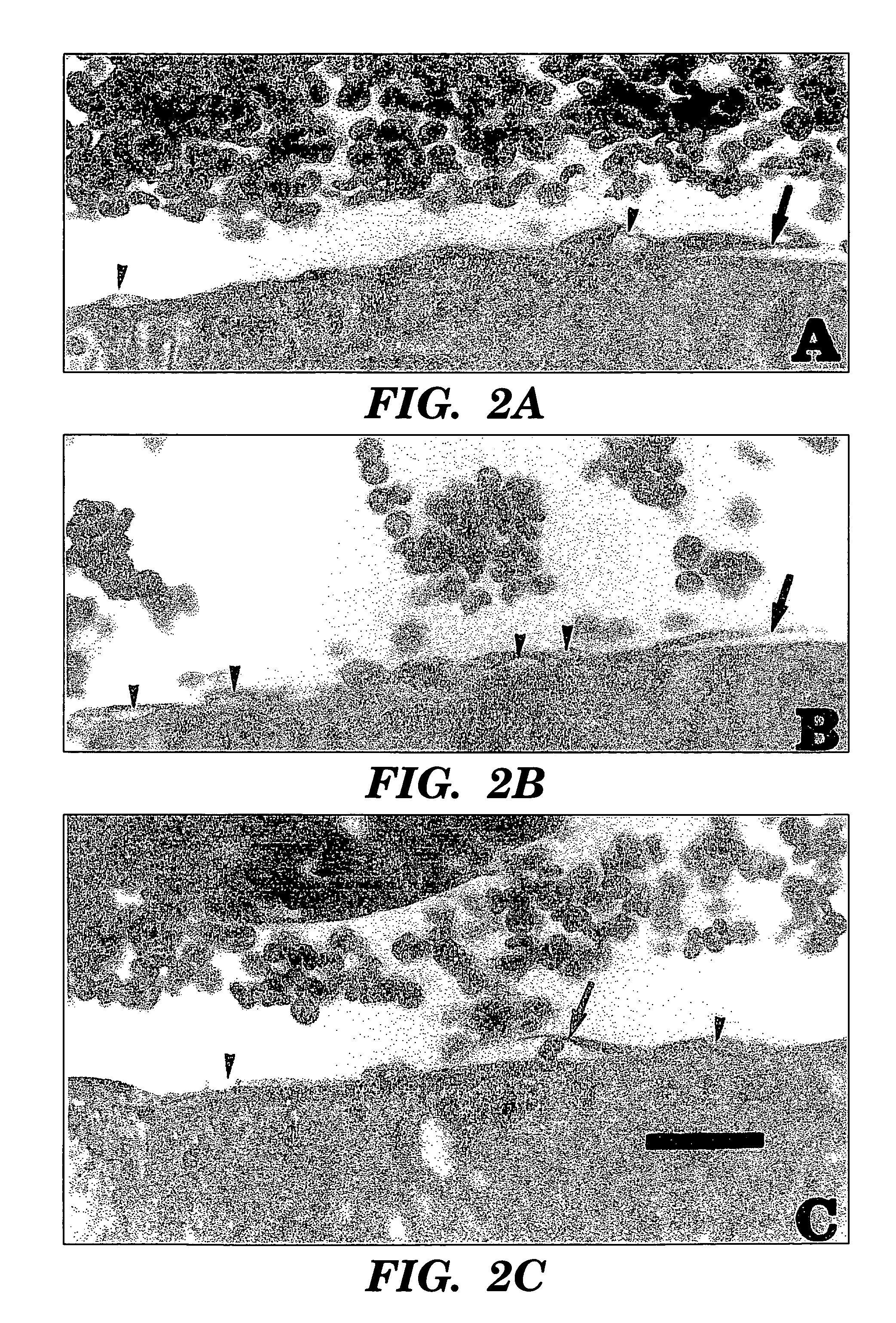
[0043]US application can cause heating, and temperature was monitored using a thermocouple placed adjacent to the thrombosed vessel or at the surface of the femur. With application of ultrasound the average initial temperature increase at the femoral artery was 0.02° C. / min, and it was 0.04° C. / min at the surface of the femur. The maximum temperature increase after 60 min was 1.6±1.3° C. at the femoral artery and 1.1±0.7° C. at the femur. Histologically, examination showed that vessels exposed to US, regardless of other treatment components (streptokinase, ligation, clot), had a pronounced tendency for endothelial cell vacuolization, and some cells lifted up off the underlying basement membrane (FIG. 2). Occasionally, erythrocytes were seen in direct contact with the basement membrane.
PUM
 Abstract
Description
Claims
Application Information
 - R&D
- Intellectual Property
- Life Sciences
- Materials
- Tech Scout
- Unparalleled Data Quality
- Higher Quality Content
- 60% Fewer Hallucinations
Browse by: Latest US Patents, China's latest patents, Technical Efficacy Thesaurus, Application Domain, Technology Topic, Popular Technical Reports.
© 2025 PatSnap. All rights reserved.Legal|Privacy policy|Modern Slavery Act Transparency Statement|Sitemap|About US| Contact US: help@patsnap.com


