
Method and apparatus for determining alveolar opening and closing

Inactive Publication Date: 2006-10-17
TIMPEL MEDICAL BV
View PDF13 Cites 20 Cited by
  • Summary
  • Abstract
  • Description
  • Claims
  • Application Information

AI Technical Summary

Benefits of technology

[0013]In contrast to computer tomography and magnetic resonance tomography, the process according to the invention can also be carried out at the bed of the patient, because no costly instruments are necessary. In this case there is no radiation stress either for the patient or for the staff. In the case of critical patients constant supervision of the state and degree of openness of the lung can therefore be carried out.
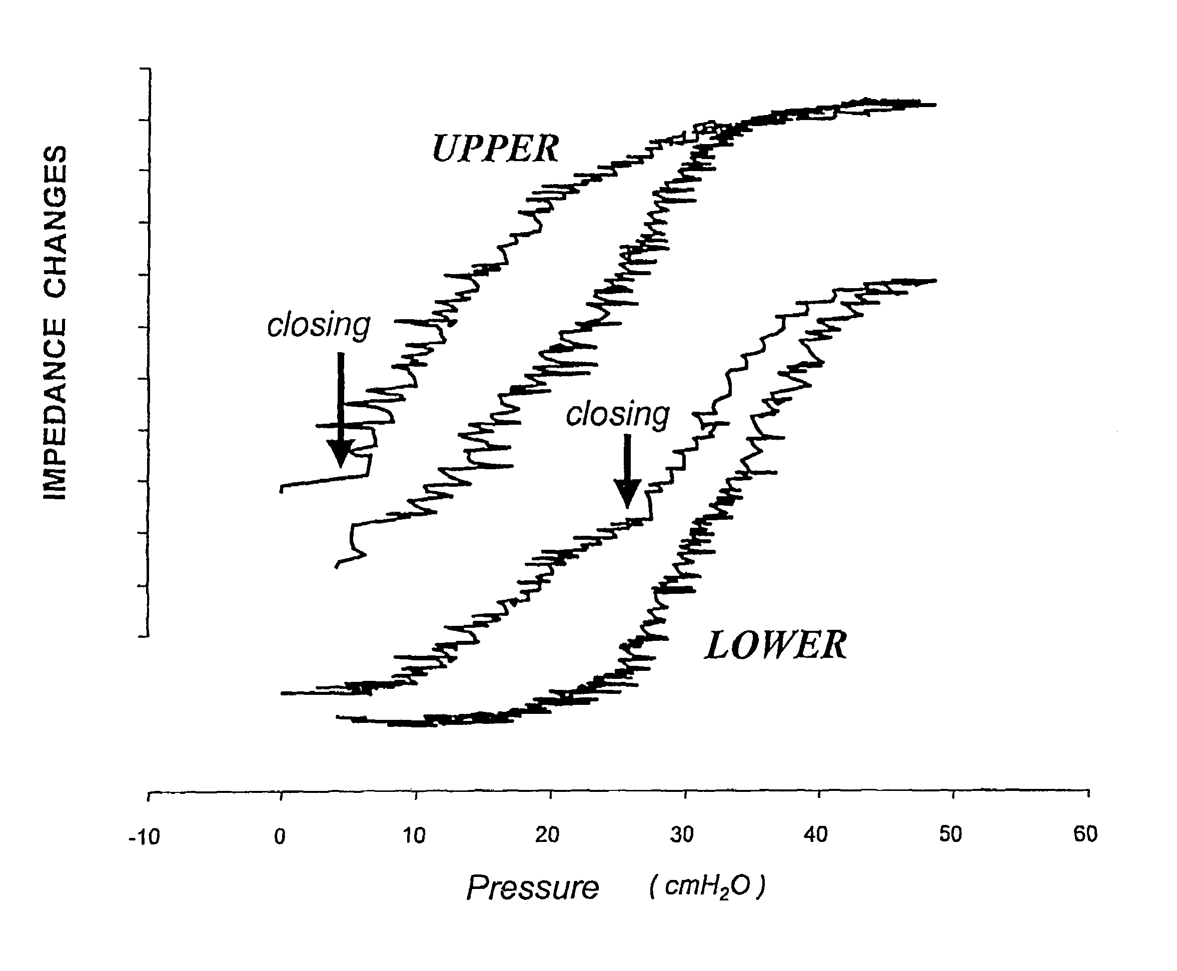
[0028]In addition, an optimal level of PEEP prevents the collapse of airways. If airways are kept open during the entire respiratory cycle, the respiratory gases are exchanged efficiently. These parts are thus ventilated and the impedance signals follow this ventilation. If, however, the conducting airways are collapsed during the entire respiratory cycle, the terminal lung units—in particular the alveoli—are cut off from the supply of fresh gas. Gas exchange suffers and no ventilation-induced change in the impedance signal can be detected. These lung areas become silent on the impedance tomographic image. The oxygen within the cut-off alveoli is absorbed and with the progressive decrease in their gas content, the absolute impedance of such a lung unit is reduced. In a scenario where PEEP levels are not high enough to prevent the expiratory collapse of airways and terminal lung units (alveoli) but where pressures are sufficiently high to open collapsed airways during inspiration, ventilation of these lung units takes place only during this period of the respiratory cycle. The changes in the impedance signals of such a lung region can be amplified compared to an area of normal ventilation since these collapsed lung units start from a low expiratory air content but are filled rapidly to approximately normal volumes during inspiration. During expiration they collapse, again and the process of tidal recruitment / collapse begins anew.
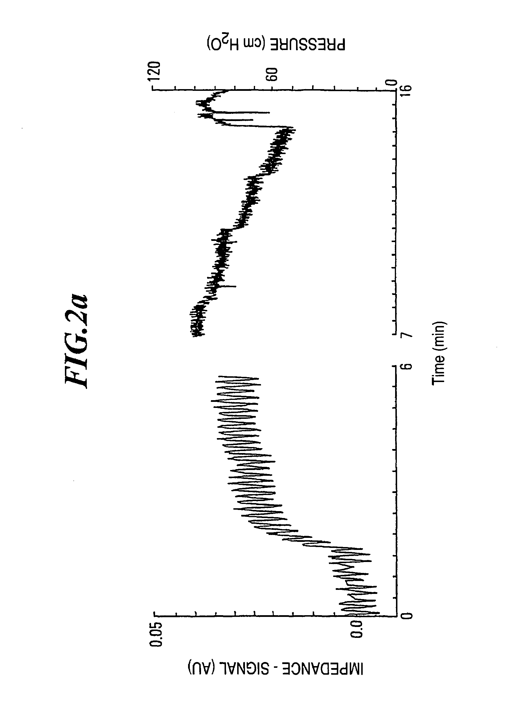
[0030]In accordance with a further aspect of the present invention, the apparatus comprises a monitoring unit for monitoring the first respiration pressure value and the second respiration pressure value. By monitoring these values the patient can be observed by a monitoring device gaining important pieces of information with regard to the lung functioning. All the direct and derived impedance signals and / or images discussed above should be calculated continuously and should be available for on-line display. Any single one of them or a combination of them can be used for the automatic or semi-automatic control of a therapeutic device, such as a mechanical ventilator. The information obtained by electrical impedance tomography can be used to guide specific clinical maneuvers aiming at optimal lung recruitment and at keeping most alveoli open or at finding the best biological compromise between alveolar over-distension and alveolar collapse.

Problems solved by technology

The artificial respiration of a sick lung, wherein oedemas have formed, is a special problem, because it cannot be exactly controlled whether the lung has already closed and / or collapsed in certain parts.
However, this pressure is not known in the case of artificial respiration, because the alveolar opening and / or closing of the lung could not yet be regionally determined.
However, in this case disturbance factors, e.g. based on offset changes, enter fully into the measurement.

Method used


Image

  • Method and apparatus for determining alveolar opening and closing
  • Method and apparatus for determining alveolar opening and closing
  • Method and apparatus for determining alveolar opening and closing

Examples


Embodiment Construction

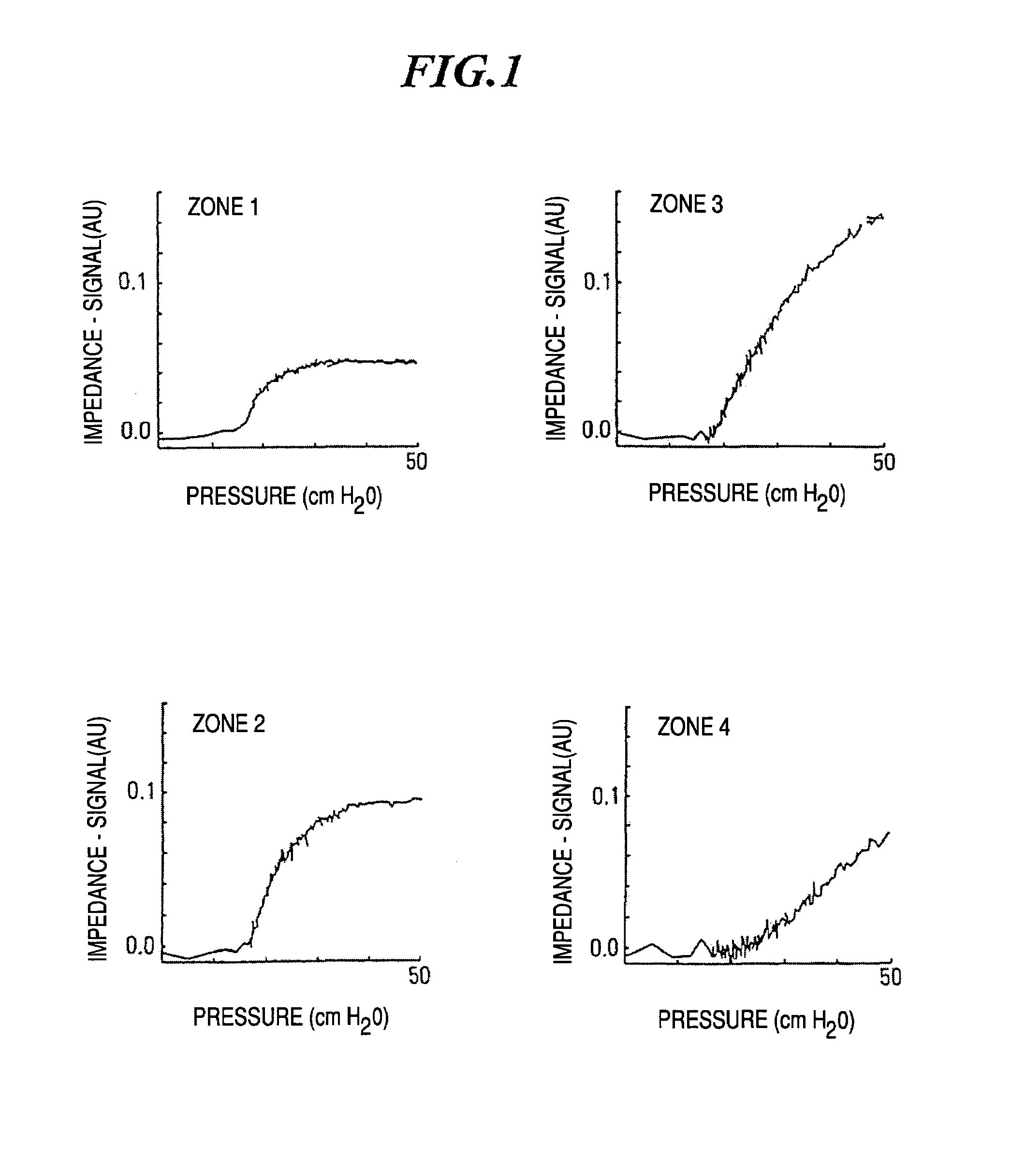
[0055]FIG. 1 shows pressure-impedance curves according to electrical impedance tomography in four different zones of the lung. In comparison with the known pressure-volume curves, the corresponding pressure-impedance curves show a similar course. As from a certain pressure point, the first alveoli (terminal lung units or air sacks) change over from the state of collapse to the state of opening. When the pressure is further increased, more and more closed alveoli are opened until the opening finally ebbs away and at higher pressures forms the flat part of the impedance signal. Comparison of the individual curves over the various zones of the lung shows that the opening phenomenon is not homogeneously distributed over the entire lung in this case. The measurements are carried out according to the method of electrical impedance tomography, wherein the zones 1 to 4 in the direction of the gravity vector subdivide the lung into planes which are perpendicular thereto. In the uppermost zon...


PUM


Abstract

The invention refers to a method for the regional determination of the alveolar opening and alveolar closing of the lung depending on the respiration pressure, wherein according to the method of electrical impedance tomography, an impedance signal is measured in at least one lung zone depending on the respiration pressure. The alveolar opening or closing of a lung zone is determined, in particular to enable an improved artificial respiration.

Description

RELATED APPLICATIONS[0001]This is a continuation of application Ser. No. 09 / 875,262, filed Jun. 7, 2001 now abandoned, which is a continuation application of application PCT / EP99 / 09699, filed Dec. 9, 1999, which claims the priority benefit of German Application No. DE 19857090.2, filed Dec. 10, 1998, which is hereby incorporated by reference.TECHNICAL FIELD OF THE INVENTION[0002]The invention refers to a method for the determination of the alveolar opening and alveolar closing of the lung depending on the pressure respiration. In particular, the invention enables to a regional determination of the alveolar opening and alveolar closing.[0003]It is known that to measure the lung mechanics, pressure and volume should be recorded and superimposed. If one increases the pressure continuously, as from a certain pressure the first alveoli (terminal lung units or air sacks) begin to change over from the state of collapse into the state of openness. If the pressure is increased further, more ...

Claims


Application Information

Patent Timeline
IPC IPC(8): A61B5/08A61M16/00A61B5/05A61B5/053A61B5/085A61B5/11
CPCA61B5/0536A61B5/08A61B5/085A61B5/0809
Inventor BÖHM, STEPHANAMATO, MARCELO B. P.KUNST, PETER W. A.
Owner TIMPEL MEDICAL BV
Learn More