
Converting diploidy to haploidy for genetic diagnosis

a technology of genetic diagnosis and haploidy, applied in the field of haploidy conversion to haploidy for genetic diagnosis, can solve the problems of complicated interpretation of sequencing ladders, affecting the accuracy of sequencing results, so as to simplify the analysis of protein products

Inactive Publication Date: 2006-05-16
THE JOHN HOPKINS UNIV SCHOOL OF MEDICINE
View PDF6 Cites 1 Cited by
  • Summary
  • Abstract
  • Description
  • Claims
  • Application Information

AI Technical Summary

Benefits of technology

[0023]Fused hybrid cells can be selected using any markers which result in a positively selectable phenotype. These include antibiotic resistance genes, toxic metabolite resistance genes, prototrophic markers, etc. The surprising advantage of the present invention is that a single marker on a single human or other mammalian chromosome can be used in the selection, and that stable hybrids containing more than just the single, selected human or other mammalian chromosome result. Thus markers on other chromosomes can be analyzed even when the chromosomes on which the markers reside were not selected.
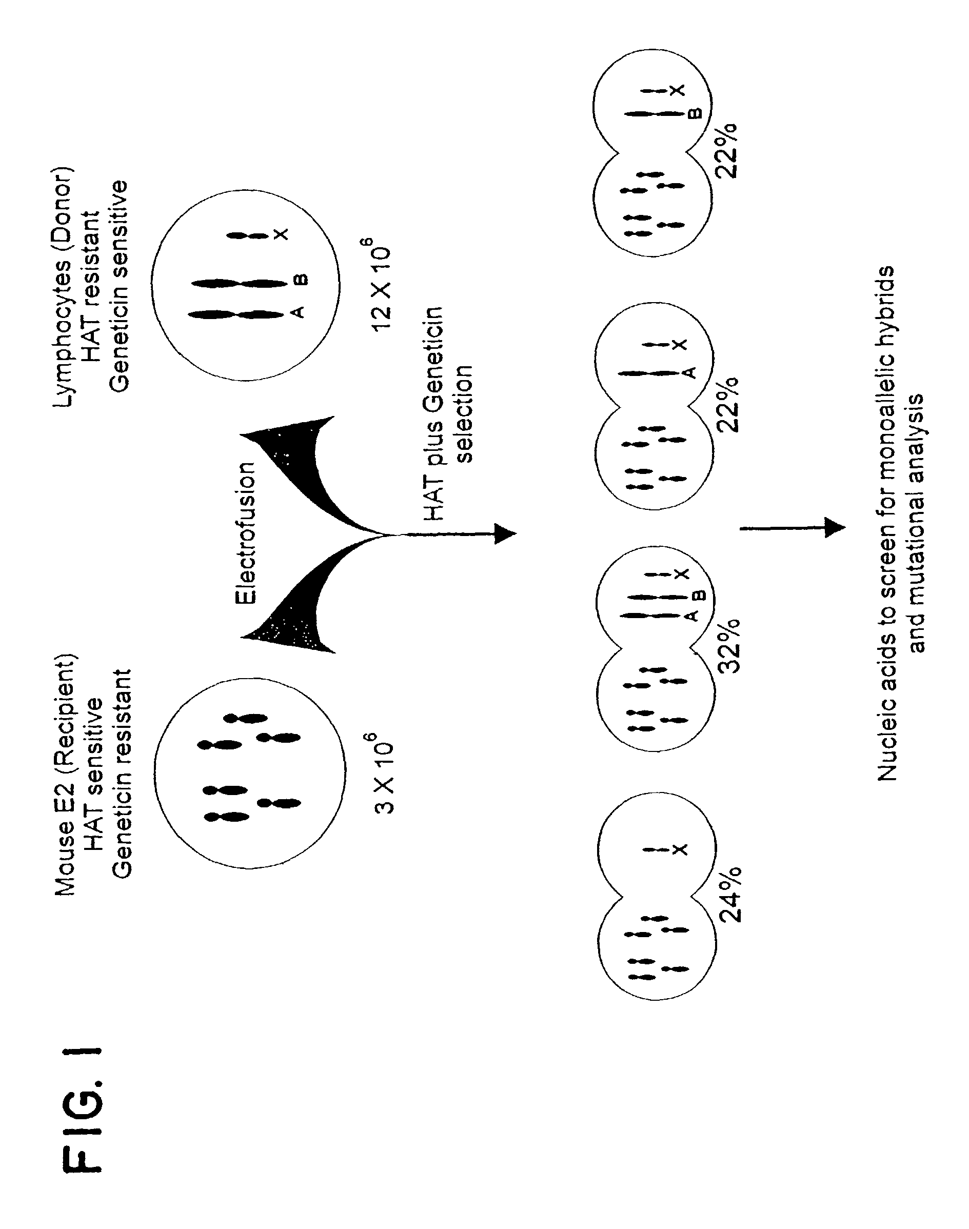
[0024]Fused hybrid cells can be analyzed to determine that they do in fact carry a human or other mammalian (non-rodent) chromosome which carries a gene of interest. Hybrid cells which have either of the two relevant human or other mammalian chromosomes can be distinguished from each other as well as from hybrids which contain both of the two human or other mammalian chromosomes. See FIG. 1. While any means known in the art for identifying the human or other mammalian chromosomes can be used, a facile analysis can be performed by assessing microsatellite markers on the human or other mammalian chromosome. Other linked polymorphic markers can be used to identify a desired human or other mammalian chromosome in the hybrids.
[0025]Once hybrid cells are isolated which contain one copy of a human or other mammalian gene of interest from a human or other mammal who is being tested, mutation analysis can be performed on the hybrid cells. The genes can be tested directly for mutations, or alternatively the mRNA or protein products of the genes can be tested. Mutations that result in reduced expression of the full-length gene product should be detectable by Western blotting using appropriate antibodies. Tests which rely on the function of the protein encoded by the gene of interest and enzyme assays can also be performed to detect mutations. Other immunological techniques can also be employed, as are known in the art.
[0026]If an immunological method is used to detect the protein product of the gene of interest in the hybrids, it is desirable that antibodies be used that do not cross-react with rodent proteins. Alternatively, the rodent genes which are homologous to the gene of interest can be inactivated by mutation to simplify the analysis of protein products. Such mutations can be achieved by targeted mutagenesis methods, as is well known in the art.
[0027]Functional tests can also be used to assess the normalcy of each allelic product. For example, if one inserted an expression construct comprising a β-galactosidase gene downstream from a p53 transcriptional activation site, into a rodent-human hybrid cell that contained human chromosome 17 but no endogenous p53, then one could detect mutations of the p53 on the human chromosome 17 by staining clones with X-gal. Other enzymatic or functional assays can be designed specifically tailored to the gene of interest.
[0028]Any method of detecting mutations at the DNA or RNA level as are known in the art may be employed. These include without limitation, sequencing, allele-specific PCR, allele-specific hybridization, microarrays, DGGE, and automated sequencing.

Problems solved by technology

The problem with humans and other mammals, at least from a genetic diagnostic perspective, is that they are diploid.
Though many powerful techniques for genetic diagnosis have been developed over the past decade, all are compromised by the presence of diploidy in the template.
For example, the presence of a wild-type band of the same electrophoretic mobility as a mutant band can complicate interpretation of sequencing ladders, especially when the mutant band is of lower intensity.
Deletions of a segment of DNA are even more problematic, as in such cases only the wild-type allele is amplified and analyzed by standard techniques.
These issues present difficulties for the diagnosis of monogenic diseases and are even more problematic for multigenic diseases, where causative mutations can occur in any of several different genes.

Method used


Image

  • Converting diploidy to haploidy for genetic diagnosis
  • Converting diploidy to haploidy for genetic diagnosis
  • Converting diploidy to haploidy for genetic diagnosis

Examples


example 1

[0034]An outline of the approach is presented in FIG. 1. The rodent fusion partner was a line derived from mouse embryonic fibroblasts transformed with ras and adenovirus E1A oncogenes. HPRT-deficient subclones of this line were generated, and one subclone (E2) was chosen for further experimentation based on its robust growth characteristics, maintenance of diploidy, and fusion efficiency (10). Human lymphocytes cells were mixed with E2 cells at an optimum ratio and electrofused, and hybrids selected in geneticin (to kill unfused human cells) and HAT (to kill unfused E2 cells) (11). Colonies appearing after two weeks of growth were expanded and RNA and DNA prepared for analysis. From a single fusion experiment, an average of 36 hybrid clones were obtained (range of 17 to 80 in five different individuals).

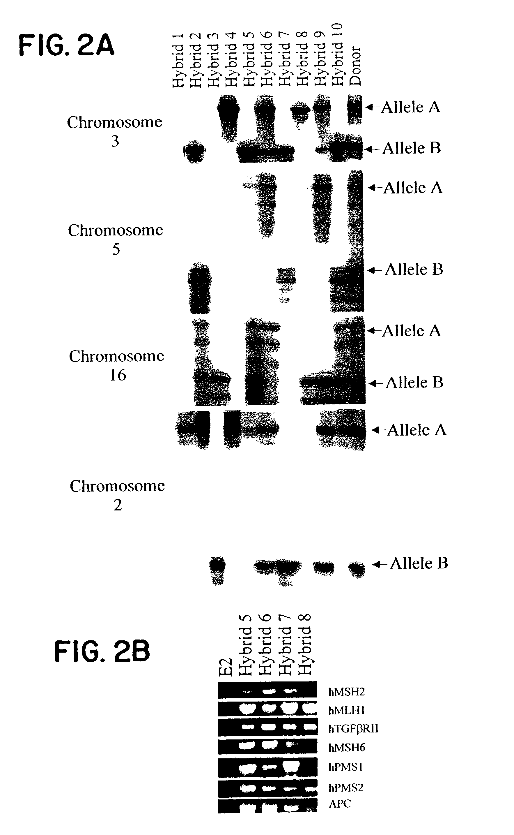
[0035]All hybrids contained the human X chromosome, as this chromosome contains the HPRT gene allowing growth in HAT. To determine whether other human chromosomes were present in th...

example 2

[0036]Two other features of the hybrids were essential for the analyses described below. First, the human chromosome complements of the hybrids were remarkably stable. Polymorphic marker analysis in ten hybrids revealed identical patterns of retention after growth for 90 (30 passages) generations after initial genotyping. Second, those hybrids containing the relevant chromosome expressed every human gene assessed, including all known colorectal cancer susceptibility genes (the hMSH2 and hMSH6 genes on chromosome 2p, the hPMS1 gene on chromosome 2q, the TGF-β Receptor Type II gene and hMLH1 gene on chromosome 3p, the APC gene on chromosome 5q, the hPMS2 gene on chromosome 7q, and the E-cadherin gene on chromosome 16q; representative examples in FIG. 2B) (14).

example 3

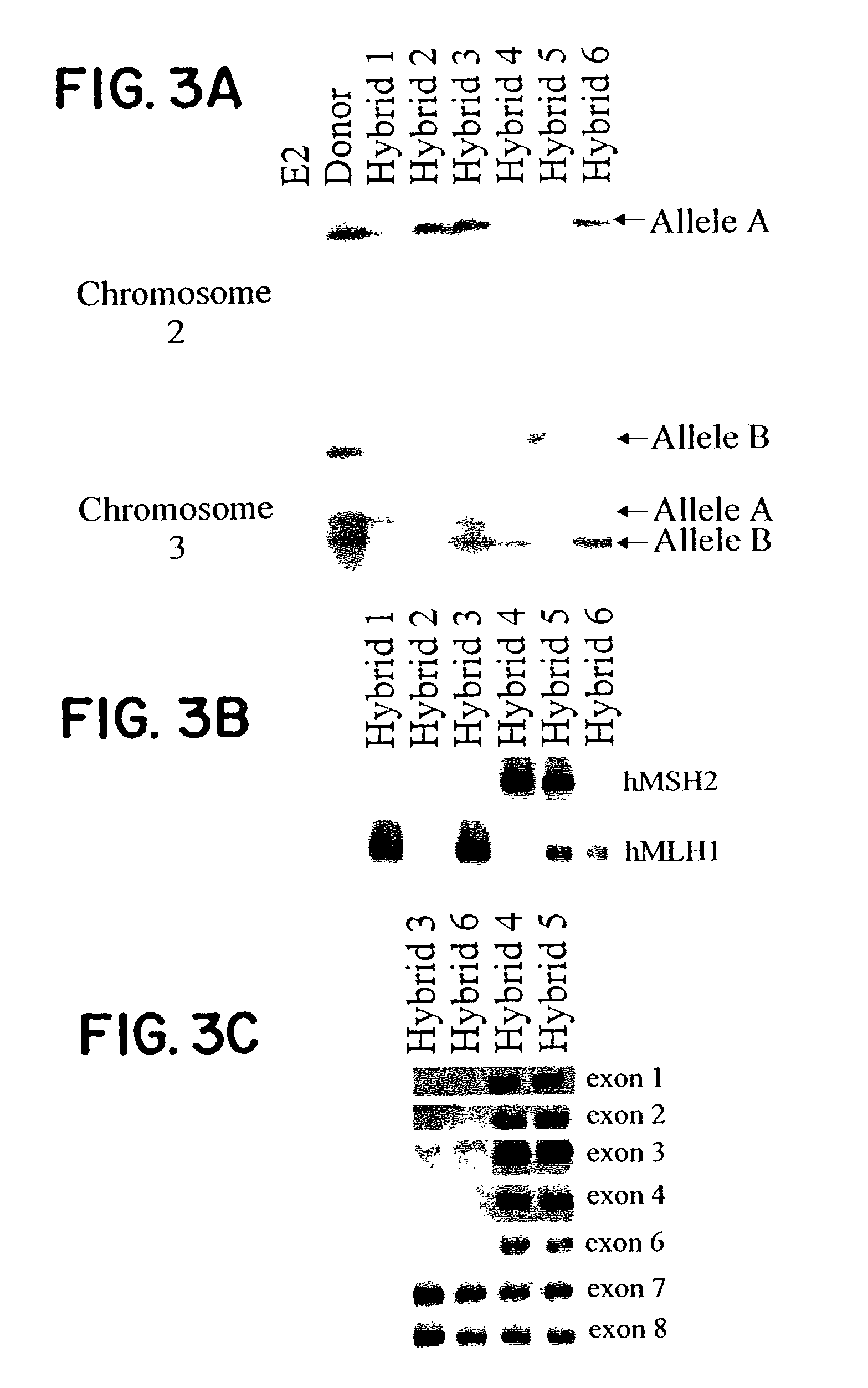
[0037]Having established the stability and expression patterns of CRC-predisposition genes in these hybrids, we used this “conversion” approach to investigate ten patients who had proven refractory to standard genetic diagnostic techniques. Each of these patients had a significant family history of colorectal cancer and evidence of mismatch repair deficiency in their tumors, yet sequencing of the entire coding sequence of each known MMR gene had failed to reveal mutations. Indeed, these and similar studies have prompted the speculation that other major HNPCC genes must exist. (25–34) Hybrids were generated from lymphocytes of each patient, and at least one hybrid containing the maternal allele and one hybrid containing the paternal allele of each MMR gene was isolated. Analysis of the nucleic acids from these hybrids revealed specific mutations in all ten patients (Table 1). In every case, an abnormality was found in a single allele of either hMSH2 or hMLH1. The nature of the abnorm...


PUM

PropertyMeasurementUnit
Fractionaaaaaaaaaa

Abstract

Detection of mutations associated with hereditary diseases is complicated by the diploid nature of mammalian cells. Mutations present in one allele are often masked by the wild-type sequence of the other allele. Individual alleles can be isolated from every chromosome within somatic cell hybrids generated from a single fusion. Nucleic acids from the hybrids can be analyzed for mutations in an unambiguous manner. This approach was used to detect two cancer-causing mutations that had previously defied genetic diagnosis. One of the families studied, Warthin Family G, was the first kindred with a hereditary colon cancer syndrome described in the biomedical literature.

Description

[0001]This is a Divisional Application of parent application Ser. No. 09 / 504,860, filed Feb. 16, 2000, now U.S. Pat. No. 6,475,794, which is a continuation-in-part of application Ser. No. 09 / 461,047 filed Dec. 15, 1999, now U.S. Pat. No. 6,399,374, which claims the benefit of provisional application Ser. No. 60 / 158,160 filed Oct. 8, 1999.[0002]This invention was supported with U.S. government funds, NIH grants CA43460, CA57345, CA62924, CA67409, CA72851. The government therefore.BACKGROUND OF THE INVENTION[0003]The problem with humans and other mammals, at least from a genetic diagnostic perspective, is that they are diploid. Mutations in one allele, such as those responsible for all dominantly inherited syndromes, are always accompanied by the wild-type sequence of the second allele. Though many powerful techniques for genetic diagnosis have been developed over the past decade, all are compromised by the presence of diploidy in the template. For example, the presence of a wild-type...

Claims


Application Information

Patent Timeline
IPC IPC(8): C12N5/12C12N15/06C12N15/87C12Q1/00G01N33/483C12N5/10C12N5/16C12N5/26C12N15/09C12Q1/02C12Q1/68C12R1/91G01N27/447G01N33/53G01N33/566G01N37/00
CPCC12Q1/6886C12N5/166C12Q2600/156C12Q1/68
Inventor VOGELSTEIN, BERTKINZLER, KENNETH W.PAPADOPOULOS, NICKOLASYAN, HAI
Owner THE JOHN HOPKINS UNIV SCHOOL OF MEDICINE
Learn More