
Surgical repair of tissue defects

a tissue defect and surgical technology, applied in the field of surgical devices and procedures, can solve the problems of increasing the duration of surgery, affecting the quality of surgical work, so as to reduce the viscosity or viscosity, and minimize the shifting of the prosthesis

Inactive Publication Date: 2006-05-16
PROMETHEAN SURGICAL DEVICES
View PDF85 Cites 93 Cited by
  • Summary
  • Abstract
  • Description
  • Claims
  • Application Information

AI Technical Summary

Benefits of technology

[0051]The invention may also include a tissue defect repair patch for implanting within a patient, comprising at least one first layer of inert synthetic mesh material sized and shaped to extend across and beyond a tissue aperture in a patient; a resilient support member adjacent a periphery of the first layer for creating tension in the first layer, the support member being carried by the first layer so as to remain implanted with the first layer in the patient; the first layer of inert synthetic mesh material having a periphery extending beyond the support member to define a border which has a free outer edge to fill uneven voids in a patient's tissue. The first layer of inert synthetic mesh material having a surgical adhesive impregnated between interstices of the first layer to effect a bond between mesh and tissue. The invention includes a tissue defect repair patch for implanting within a patient, comprising: at least one layer of flexible, inert synthetic non-porous sheet sized and shaped to extend across and beyond a tissue aperture in a patient; a resilient support member adjacent a periphery of the layer for creating tension in the layer, the support member being carried by the layer so as to remain implanted with the layer in the patient; and the layer of inert synthetic sheet having a periphery extending beyond the support member, defining a border which has a free outer edge to fill uneven voids in a patient's tissue; and the layer of inert synthetic sheet having a surgical adhesive coating the sheet to effect a bond between mesh and tissue. The sheet may be comprised of a first substance encapsulating a surgical adhesive, such that when pressure is applied, the encapsulated surgical adhesive is ejected from the encapsulation sites to effect a bond between the ruptured cell structure and tissue. The patch may have a plurality of slits in the border. The invention may comprise a plug formed of non-porous material in which a central cavity allows for delivery of adhesive; a

Problems solved by technology

Polypropylene is used because it promotes a fibroblastic response, but this response is only important for ensuring mesh fixation, it is also considered to be too stiff.
The need to shape the mesh during the surgery increases the duration of the surgery and also makes optimal fitting difficult.
Suturing requires a great deal of skill, especially for minimally invasive or laparoscopic procedures.
The difficulty in placing sutures is a primary barrier to the general adoption of laparoscopic procedures.
Both the suture and staple approaches risk a variety of complications.
Frequently, because of possible excessive tension and bad distribution of the prosthesis, it isn't able to dynamically suit the modification of relationships among the anatomic structures of the inguinal canal and because it is suture along its periphery.
This produces pain that can be felt during movements of the muscular-aponeurotic structures in the inguinal region.
The sutures themselves can cause vascular lesions, tissue lacerations, and nerve lesions.
Plugs however, have their own problems.
Perception of an inguinal tumefaction connected to sclerosis stimulated by the presence of the plug is experienced as discomfort by the patient.
Relapses after using the plug are difficult to treat.
There is difficulty in treating postoperative suppuaration (formation of pus) because the presence of the underfascial plug cannot be cleaned with superficial dressing.
Given the hernia rate in the United States population, the constant use of properitoneal underfascial plugs could cause a significant amount of risk to patients because of possible prosthetic infection if an operation were necessary for a pelvic septic pathology such as tumor of the colon, appendicitis, or surgeries of the prostate and bladder.
The plug also tends to displace on the operating table, forcing surgeons to secure the mesh with stitches, thus undermining the sutureless advantage.
Finally, if the sutureless technique is used in the case of an external oblique inguinal hernia, the yielding of the transversalis layer is coupled with the yielding of the external inguinal ring and of the aponeurosis of the great oblique, said aponeurosis sometimes becomes a fibrous, sliding veil unsuitable to support the prosthesis under abdominal pressure.
The method risks tissue damage from the energy required to be deposited in the tissue to bond collagen pads to living tissue.
This implant is heat molded and stiff, and does not conform to irregularities in the muscle or tissue wall defining the defect.
However, there is provided no means for fixing the implant position.
With respect to endoscopic techniques, concerns have been raised about visualization light reflected from the fabric surface during laparoscopy, impairing visualization of the prosthetic material and underlying anatomy.
Increasing the pore size of the mesh helps, but also diminishes the physical properties of the implant necessary for augmentation or repairing abdominal tissue.

Method used


Image

  • Surgical repair of tissue defects
  • Surgical repair of tissue defects
  • Surgical repair of tissue defects

Examples


Embodiment Construction

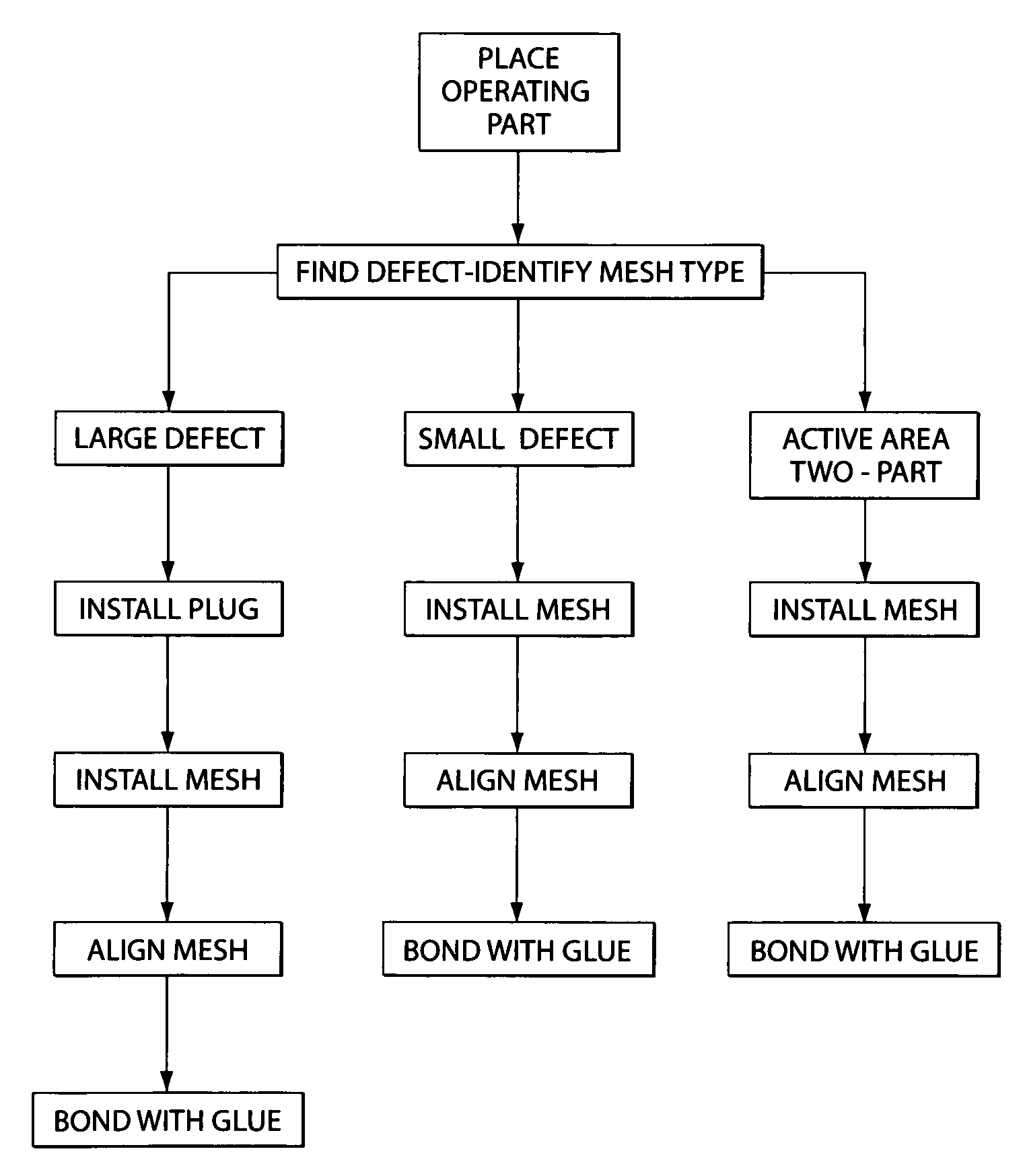
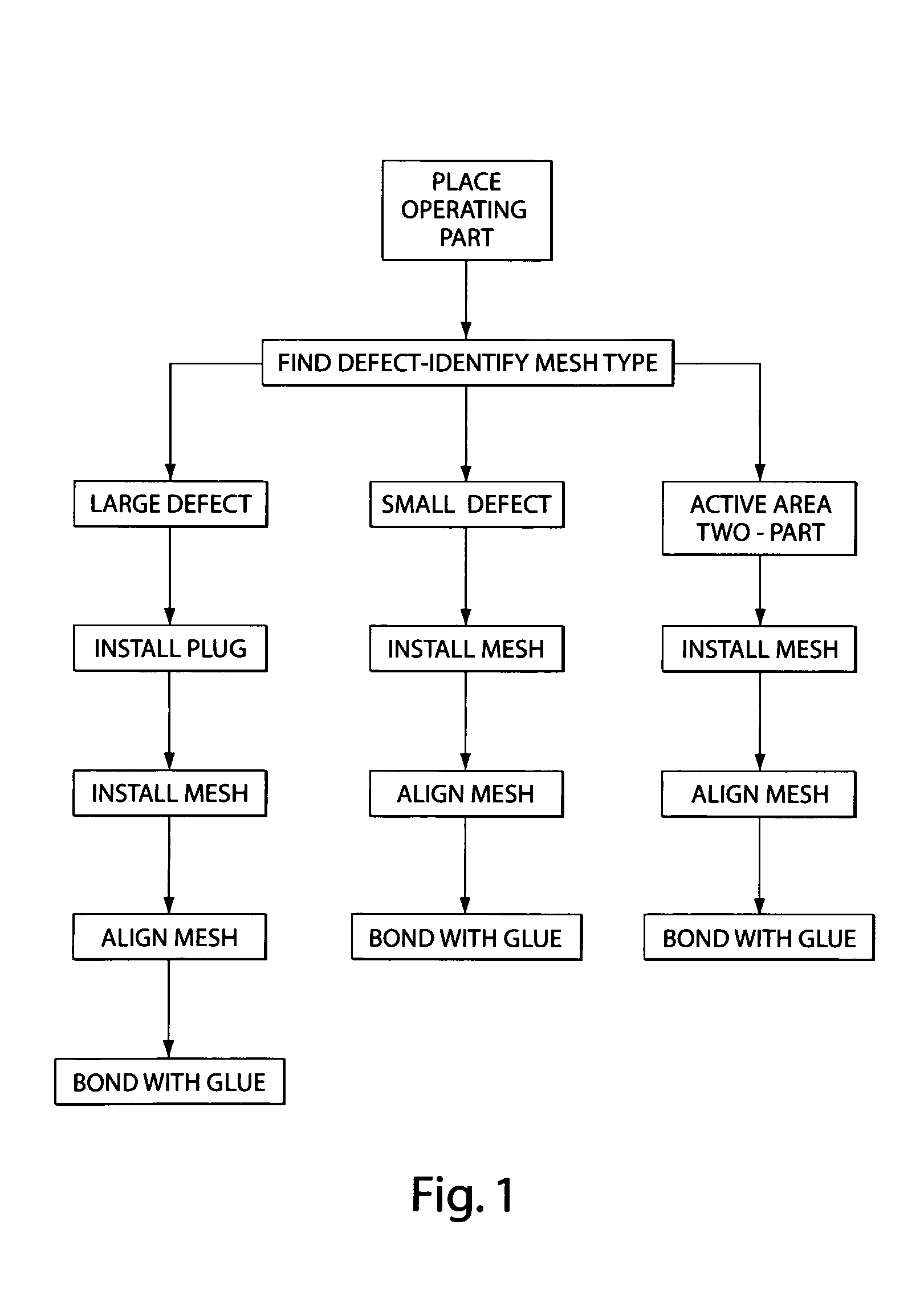
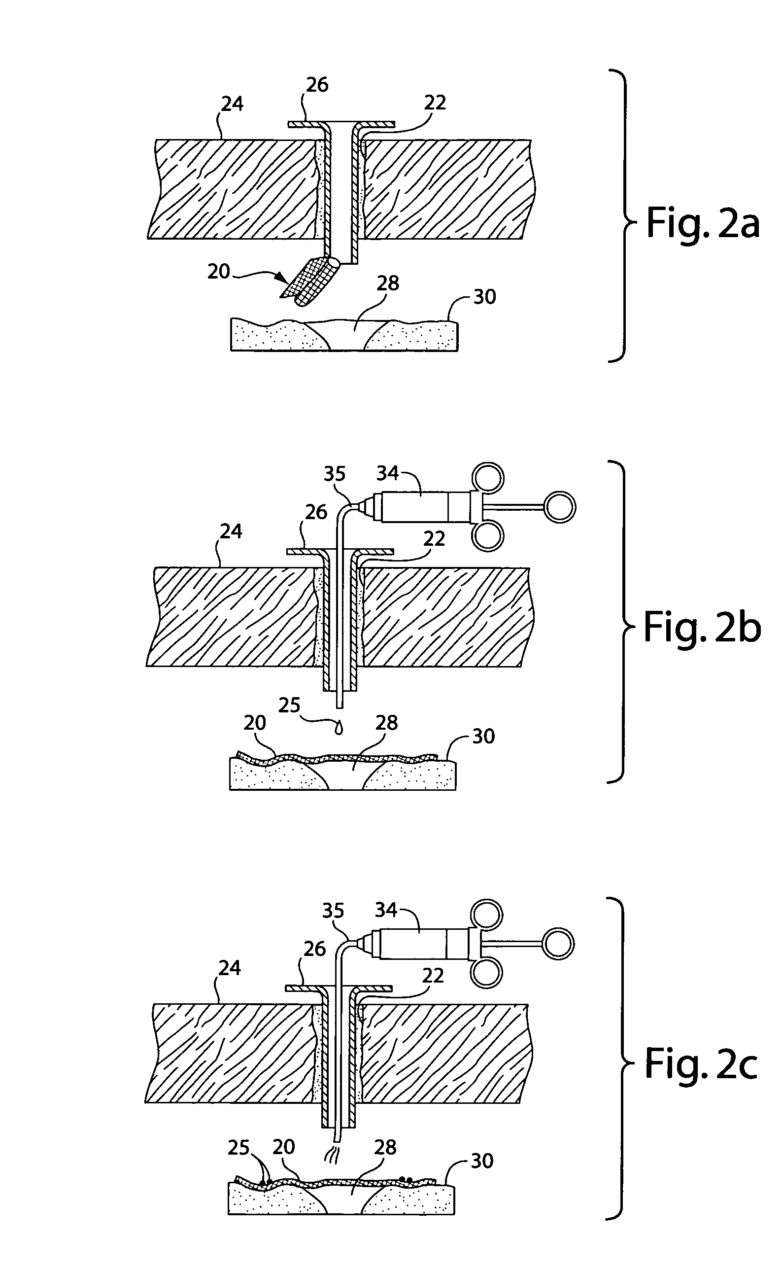
[0061]Referring now to the drawings in detail, and particularly to FIG. 1, there is shown in a block diagram format, the process of the present invention, and in FIGS. 2a, 2b and 2c, as schematic representations of the surgical repair apparatus and its associated procedure for treating tissue defects.

[0062]The invention thus comprises a mesh 20 intended to be implanted laparoscopically, as may be envisioned in FIGS. 2a–2c, and constructed so as to allow for it to be delivered through a small hole or access port 22 in the skin 24. Meshes 20 delivered in this way are preferably rolled into a cylinder, as shown in FIG. 2a, passed through a trocar 26 in the access port and unfurled over the tissue defect 28. Additional instrumentation may be introduced in the access port 22 for the purpose of suturing or stapling the mesh 20 to the tissue 30. One of the complications of introducing an adhesive coated mesh in this way is the likelihood that it will adhere to itself or to surrounding tiss...


PUM

PropertyMeasurementUnit
Solubility (mass)aaaaaaaaaa
Water solubilityaaaaaaaaaa
Sensitivityaaaaaaaaaa

Abstract

A method for repairing a defect in living mammalian tissue comprising: covering a tissue defect and surrounding tissue with a prosthetic by placing the prosthetic over the defect and against the surrounding tissue. The method includes applying a surgical adhesive to the prosthetic on the surrounding tissue on at least one location on the prosthetic and the surrounding tissue so that surrounding tissue and the prosthetic adhere to each other.

Description

[0001]This application is a continuation-in-part of U.S. Ser. No. 09 / 692,963, now U.S. Pat. No. 6,296,607, entitled “In-Situ Bulking Device”, filed Oct. 20, 2000, which is incorporated herein by reference in its entirety.BACKGROUND OF THE INVENTION[0002]1. Field of the Invention[0003]Field of the Invention. This invention relates to surgical devices and procedures, and more particularly for the repair and augmentation of mammalian tissue utilizing a combination of adhesives and prosthetics, such repair, for example treating an inguinal hernia.[0004]2. Prior Art[0005]The surgical repair treatment of inguinal hernias fall into two groups: repairs that involve suturing the prosthesis to the surrounding muscular aponeurotic structures, such as Lichtenstein's operation; and the repairs that involve application of a sutureless prosthesis such as Gilbert's operation.[0006]In the current medical practice, the established procedure in the surgical field is to use mesh, absorbable and non-abs...

Claims


Application Information

Patent Timeline
IPC IPC(8): A61F2/02A61L27/00A61F2/00A61L24/00A61L27/18A61L27/50
CPCA61F2/0036A61L27/18A61L27/50C08L75/04A61P1/04A61P13/00
Inventor MILBOCKER, MICHAEL
Owner PROMETHEAN SURGICAL DEVICES
Learn More