
Endotracheal tube

Inactive Publication Date: 2005-05-10
RGT UNIV OF CALIFORNIA
View PDF20 Cites 12 Cited by
  • Summary
  • Abstract
  • Description
  • Claims
  • Application Information

AI Technical Summary

Benefits of technology

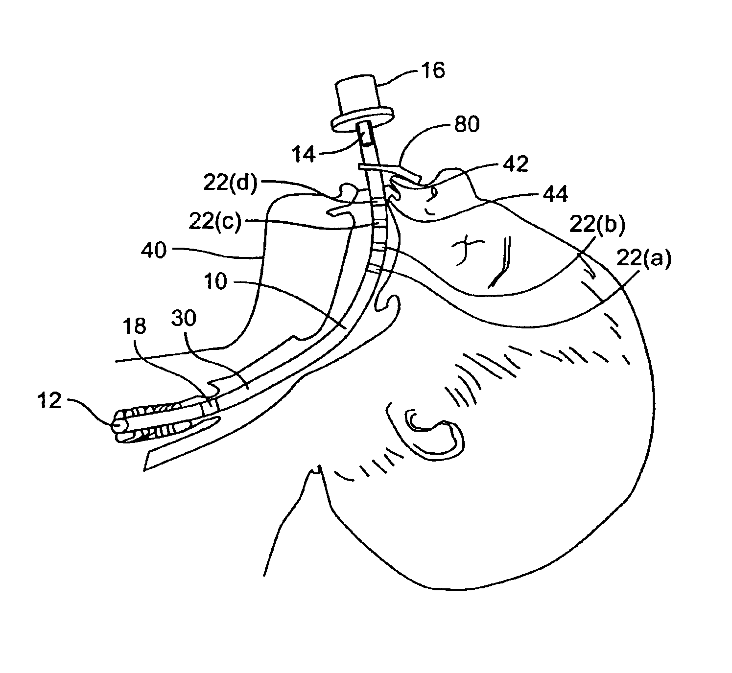
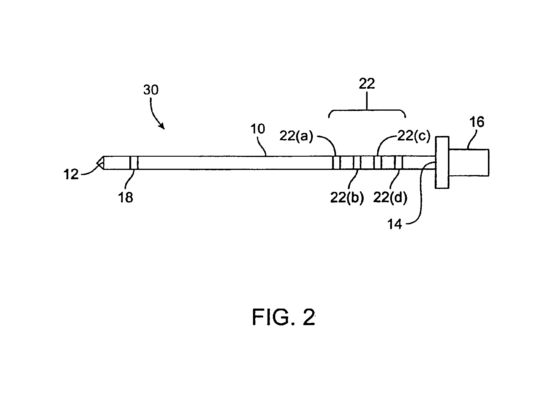
[0015]In preferred embodiments of the invention, the distinct regions on the endotracheal tube are brightly colored lines. During intubation, one of the lines is selected and localized at, for example, a fixed anatomical location such as the upper gingival ridge. The depth of insertion for the endotracheal tube can be easily determined by viewing the selected and localized line, even though the distal end of the endotracheal tube is not visible to the caregiver. Moreover, the brightly colored lines are more easily discernable to the caregiver than, for example, mono-colored markings such as black printed numbers. Accordingly, by using embodiments of the invention, the occurrence of complications from improper intubation such as RMSBI is reduced.

Problems solved by technology

The endotracheal tube made of this soft material is difficult to manipulate and insert into the trachea (windpipe).
Consequently, it is difficult to determine the ultimate depth of insertion.
Also, during prolonged periods of intubation that provide assisted breathing, it is difficult to tell if the endotracheal tube has moved from its initial, secured position.
Inserting an endotracheal tube too far into the patient can be hazardous.
For example, if an endotracheal tube extends beyond the carina (i.e., the point at which the trachea branches into the right and left lung) and into the patient's right mainstem bronchus, the bronchial intubation can result in injury.
Right mainstem bronchial intubation (RMSBI) can cause the complications listed below or even death of the patient.
RMSBI is a potential cause of morbidity during neonatal assisted ventilation (breathing).
Thus, the margin for error when inserting and maintaining an endotracheal tube in neonates is especially small.

Method used


Image

  • Endotracheal tube
  • Endotracheal tube
  • Endotracheal tube

Examples


example

[0054]The present inventors performed two studies to ascertain the incidence of RMSBI in intubated neonates. Specifically, a retrospective study and a prospective study were performed.

Retrospective Study

[0055]A first phase of this study retrospectively reviewed the incidence of RMSBI at a particular medical center. This first phase was conducted before evaluating the new endotracheal tube design according to embodiments of the invention. The retrospective review was for the years 1996, 1997, and 1998. A database identified all intubated infants. Charts of these patients were abstracted for demographic information and reports of all chest radiographs. The protocol in the NICU (neonatal intensive care unit) for performing chest radiographs kept the head in the midline position with the arms extended beside the head. During the 1996 through 1998 period, the endotracheal tube had the same localizing marks as standard neonatal endotracheal tube in use today. This endotracheal tube was us...


PUM


Abstract

An endotracheal tube and a method for using an endotracheal tube are disclosed. In one embodiment, the endotracheal tube includes a tubular member including a distal end and a proximal end. The endotracheal tube also includes a plurality of visually distinct regions that is closer to the proximal end than the distal end. Each visually distinct region is spaced from other visually distinct regions, and each of the distinct region comprises a respectively different color.

Description

CROSS-REFERENCES TO RELATED APPLICATIONS[0001]This patent application is a continuation application of U.S. patent application Ser. No.10 / 040,982, filed on Dec. 28, 2001, now U.S. Pat. No. 6,668,832, which is herein incorporated by reference in its entirety for all purposes.STATEMENT AS TO RIGHTS TO INVENTIONS MADE UNDER FEDERALLY SPONSORED RESEARCH OR DEVELOPMENT[0002]Not applicable.BACKGROUND OF THE INVENTION[0003]Endotracheal intubation is a common medical procedure. Endotracheal tubes are used in many situations. They are used as conduits for airway life support where patients have stopped breathing on their own (e.g., because of cardiac arrest, respiratory distress / arrest, seizures, and the like). They are also used in both human and animal surgery to provide passageways for anesthetic gases into lungs.[0004]Endotracheal tubes are typically made of soft biomaterials. The most common biomaterial is polyvinyl chloride with a plasticizer added, but other synthetic materials are ei...

Claims


Application Information

Patent Timeline
IPC IPC(8): A61M16/04
CPCA61M16/04A61M2205/6081A61M16/0434
Inventor HIPOLITO, RONALDO B.SHERMAN, MICHAEL P.MILSTEIN, JAY M.
Owner RGT UNIV OF CALIFORNIA
Learn More