
Systems for percutaneous bone and spinal stabilization, fixation and repair

Inactive Publication Date: 2000-10-03
KYPHON
View PDF51 Cites 1284 Cited by
  • Summary
  • Abstract
  • Description
  • Claims
  • Application Information

AI Technical Summary

Benefits of technology

It is an object of the present invention to provide devices and methods which allow intramedullar bone fixation using minimally-invasive surgical procedures, so as to generally reduce operative trauma and allow speedier post-operative recovery.
FIG. 36 is a schematic cross sectional illustration of the intramedullar nail of FIG. 35, after activation of the inflatable member to increase the diameter of the intramedullar nail, making it fit tightly against the inner surface of the bone cortex, thereby eliminating the loosening illustrated in FIG. 35.

Problems solved by technology

The problem of holding the fixture in position is complicated by the fact that the intramedullar cavity of most long bones is not uniform, but is, rather, narrow at the middle of the bone and flares out at the ends.
The problem is further complicated by the fact that a rod inserted into such canal does not normally provide stabilization for rotational and bending movement.
Such reaming destroys tissue within the bone and may consequently retard proper healing.
Such protruding portions, however, increase the likelihood of postoperative infection and interfere with mobilization of the bone.
However, a simple treatment which allows early mobilization with fracture stabilization is not currently available.
It also does not allow good rotational stabilization.
In addition, it can also result in infection due to communication between the bone medulla and the exterior of the body.
There are patents that exist today for bone fixation with a balloon, but these do not provide intramedullary nailing.
Back pain is a widespread ailment which is often attributed to disc pathologies and vertebral instability.
Patents and applications employing artificial intervertebral discs currently exist, although they have not yet been proven successful in patients (U.S. Pat. No. 4,759,769; WO 92 / 14423; WO 90 / 00037; WO 96 / 37170).
This does not allow for the insertion of the device percutaneously in a minimally invasive technique.
Such open laminectomy is a major surgical procedure, with attendant risks, side effects and long recovery time.
The procedure is successful only in about 70% of cases or less, however, and does not allow the full range of treatment afforded by open back surgery.
For example, disc prostheses and methods of implantation of such prostheses that are known in the art are not suitable for use in the percutaneous approach.
The herniated nucleus may exert pressure on a spinal nerve adjacent to the disc, resulting in pain or loss of muscle control.
The normal procedure in such cases is to remove the herniated disc tissue in open surgery, but this is a major procedure with long recovery and potentially serious side effects.
Removal of the herniated tissue is a long process, however, frequently requiring removal and reinsertion of resection tools many times.
However, percutaneous diskectomy procedures and devices known in the art do not generally achieve such thorough cleaning.
In the past, prostheses have often shown a decrease in effectiveness over time due to loosening effects.
For example, hip prosthesis implants and knee prosthesis implants have shown a high rate of implant loosening over time.
Likewise, it is known that bone implant complications generally can include loosening between the implant and the bone, causing relative movement between the bone and the implant, and forcing another operation to achieve good bone to implant fixation.
In these embodiments, the inflation of a balloon causes plastic deformation of the implant material, resulting in deformation of the device.
Rotation of the eroding element, whether the brush, the spinning wire or the orb, causes erosion of soft tissue in the eroding element's vicinity.

Method used


Image

  • Systems for percutaneous bone and spinal stabilization, fixation and repair
  • Systems for percutaneous bone and spinal stabilization, fixation and repair
  • Systems for percutaneous bone and spinal stabilization, fixation and repair

Examples


Embodiment Construction

Features of the invention are further illustrated by reference to the figures, the following description and the claims, which provide additional disclosure of the invention in various preferred embodiments.

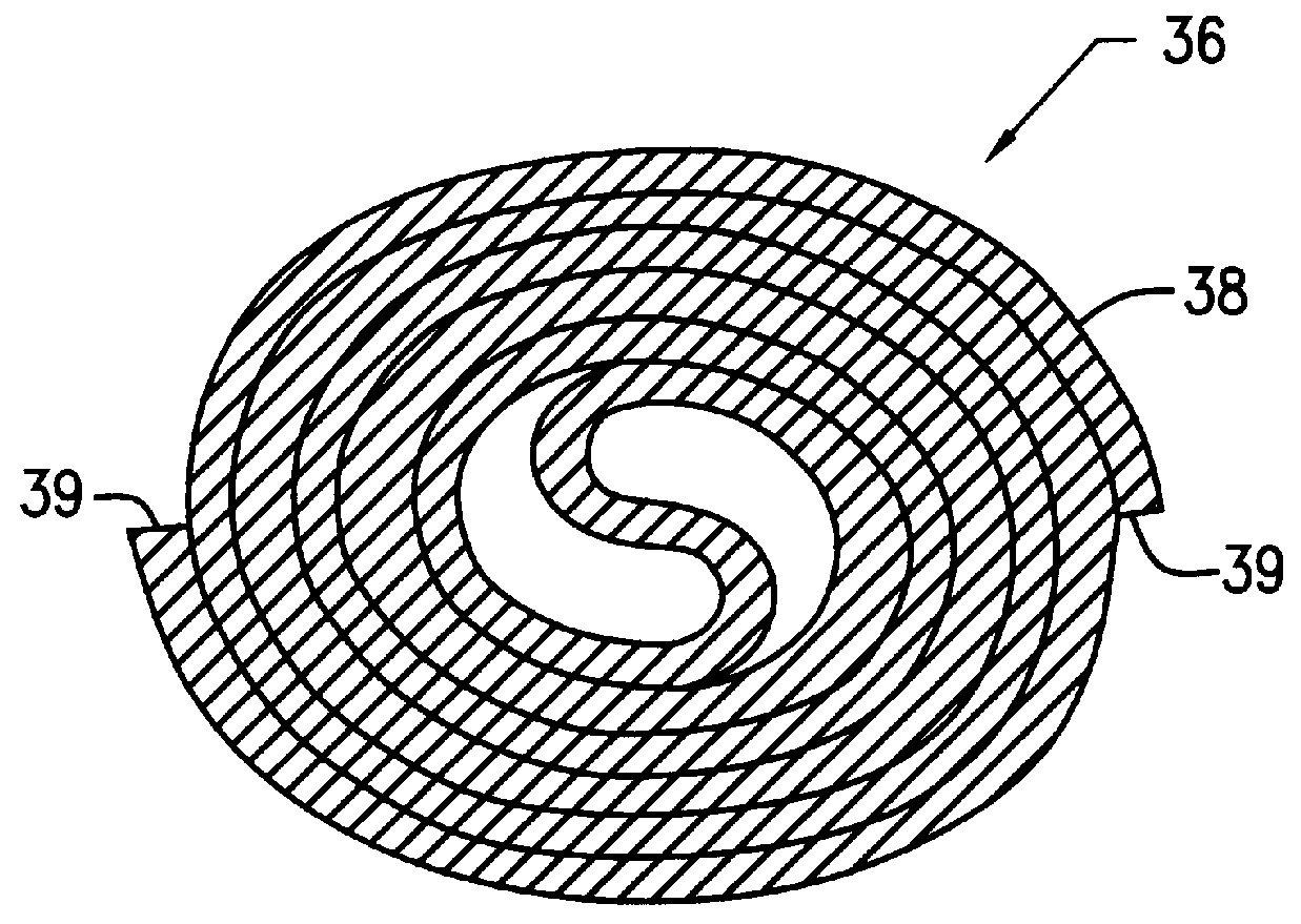
FIG. 1 is a schematic, isometric representation of a self-expanding intramedullar fixture 20, in accordance with a preferred embodiment of the present invention. Fixture 20 is preferably constructed of two sheets 22 and 24 of resilient, biocompatible material, preferably a superelastic material or a shape memory material, as is known in the art. Nitinol is preferred. Alternatively, the fixture may be constructed from another biocompatible metal, such as titanium, or a plastic or polymer material.

Sheets 22 and 24 are initially rolled tightly together into a cylindrical form. Each sheet of this compacted form is tightly rolled (as generally shown in FIG. 2A), and fixture 20 is inserted, in this compacted form, into the intramedullar cavity of a bone (FIG. 3B), as described below. W...


PUM

PropertyMeasurementUnit
Fractionaaaaaaaaaa
Flow rateaaaaaaaaaa
Diameteraaaaaaaaaa

Abstract

Systems for bone and spinal stabilization, fixation and repair include intramedullar nails, intervertebral cages and prostheses, remotely activatable prostheses, tissue extraction devices, and electrocautery probes. The intramedullar nails, intervertebral cages and prostheses, are designed for expansion from a small diameter for insertion into place to a larger diameter which stabilizes, fixates or repairs the bone, and further can be inserted percutaneously. Remotely activatable prostheses can be activated from an external unit to expand and treat prosthesis loosening. Tissue extraction devices, and electrocautery probes are used to remove tissue from desired areas.

Description

The present invention relates to systems for percutaneous bone and spinal stabilization, fixation and repair, including intramedullar bone and vertebral fixtures, vertebral disc prostheses, and electrocautery probes.BACKGROUND OF THE INVENTIONSIntramedullar Fixtures for Repair of Broken BonesIntramedullar fixtures for use in repair of broken bones are well known in the art. Such fixtures, which generally have the form of long, narrow nails, are inserted longitudinally into the bone's intramedullar cavity, so as to connect together and jointly brace two or more sections of a severely fractured bone, and thereby promote healing.A fixture of this type must have a radial diameter large enough to firmly and rigidly hold its position after insertion. The problem of holding the fixture in position is complicated by the fact that the intramedullar cavity of most long bones is not uniform, but is, rather, narrow at the middle of the bone and flares out at the ends. The problem is further com...

Claims


Application Information

Patent Timeline
IPC IPC(8): A61B17/72A61B17/68A61F2/44A61F2/46C21D9/26G01L7/00G01L7/16A61F2/00A61F2/30A61B17/00A61B17/58A61B17/03
CPCA61B17/7266G01L7/16A61F2/441A61F2/4455A61F2/446A61F2/4611C21D9/26A61B17/7275A61B17/7283A61B17/7291A61B2017/00535A61B2017/00867A61F2/442A61F2002/30093A61F2002/30098A61F2002/30293A61F2002/30579A61F2002/4627A61F2210/0019A61F2210/0033A61F2230/0091A61F2002/30092
Inventor BEYAR, MORDECHAYGLOBERMAN, ORENMAGAL, ELAD
Owner KYPHON
Learn More