
System and method for generating a stained image

a technology of system and method, applied in image generation, image enhancement, instruments, etc., can solve the problems of time-consuming and costly physical staining methods

Pending Publication Date: 2022-10-27
CITY UNIVERSITY OF HONG KONG
View PDF6 Cites 1 Cited by
  • Summary
  • Abstract
  • Description
  • Claims
  • Application Information

AI Technical Summary

Benefits of technology

The patent text describes a method for generating simulated or modelled images of tissue samples that have been stained with different stains or staining agents. These images can be used for diagnostic or medical assessments. The method utilizes multi-modal image synthesis, which allows for the combination of multiple input images to create the output image, resulting in improved accuracy. The technical effect of this method is the ability to generate images of tissue samples with different stains, increasing the flexibility and accuracy of image generation for research and diagnostic purposes.

Problems solved by technology

However, physical staining methods can be time consuming and costly.
Further, they may contribute to workplace and environmental pollution.

Method used


Image

  • System and method for generating a stained image
  • System and method for generating a stained image
  • System and method for generating a stained image

Examples


Embodiment Construction

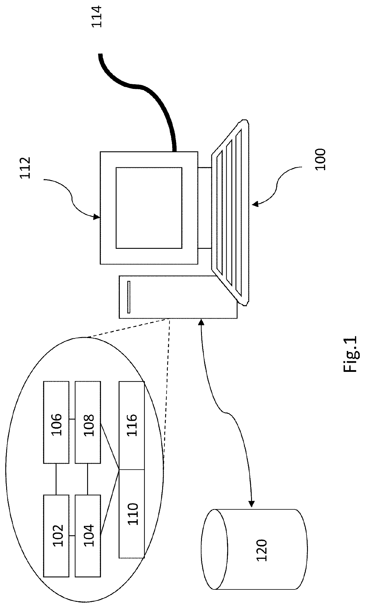
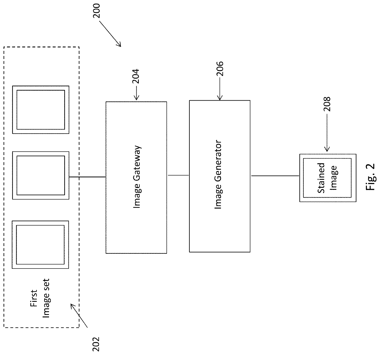
[0054]Referring to FIG. 1, an embodiment of the present invention is illustrated. This embodiment is arranged to provide a system for generating a stained image, comprising:[0055]an image gateway arranged to obtain a first image of a key sample section; and[0056]an image generator arranged to process the first image with a multi-modal stain learning engine arranged to generate at least one stained image, wherein the at least one stained image represents the key sample section stained with at least one stain.

[0057]In this example embodiment, the image gateway and image generator are implemented by a computer having an appropriate user interface, communications port and processor. The computer may be implemented by any computing architecture, including stand alone PC, client / server architecture, “dumb” terminal / mainframe architecture, portable computing devices, tablet computers, wearable devices, smart phones or any other appropriate architecture. The computing device may be appropri...


PUM

PropertyMeasurementUnit
thicknessaaaaaaaaaa
thickaaaaaaaaaa
microscopyaaaaaaaaaa

Abstract

A system and method for generating a stained image including the steps of obtaining a first image of a key sample section; and processing the first image with a multi-modal stain learning engine arranged to generate at least one stained image, wherein the at least one stained image represents the key sample section stained with at least one stain.

Description

TECHNICAL FIELD[0001]The present invention relates to a system and method for generating a stained image, and particularly, although not exclusively, to a system and method for generating a virtually stained image of a tissue sample section.BACKGROUND[0002]The practice of histology is an important area of medical sciences that has helped with medical research, diagnosis and treatment. Part of this practice involves the preparation of slides which contain tissue sections. In turn, these slides can be placed under a microscope for further study or analysis.[0003]Staining tissue for contrast when viewed under a microscope is an integral part of pathology. In various situations, a staining agent or stain, may be used to stain the tissue sections, such as by immersing tissue in chemical or antibody solutions prior to microscopy. These staining agents allow certain cells, features or structures to become more visible when the slides are viewed or considered under magnification. However, p...

Claims


Application Information

Patent Timeline
Patent Type & Authority Applications(United States)
IPC IPC(8): G06T5/00G06T5/50G06T7/30G06T3/40G01N1/30
CPCG06T5/008G06T5/50G06T5/001G06T7/30G06T3/4007G01N1/30G06T2207/10064G06T2207/20081G06T2207/30024G01N2001/302G06T11/001G06N3/08G06T2210/41G06V10/82G06V20/69G06V10/772G06V10/774G06T5/94G06T5/00
Inventor LAU, CONDONHUI, TIK HOYUAN, YIXUANCHEN, ZHENCHO, CHI SHINGCHEUK, WAHLAW, WING LUNMARASHLI, MOHAMAD ALIPANI, ANUPAMHILL, FRASER
Owner CITY UNIVERSITY OF HONG KONG
Learn More