Cancer Progression Risk Assessment by Microvascular Phenotype

a microvascular and risk assessment technology, applied in the field of cancer progression risk assessment by microvascular phenotype, can solve the problems of reducing the risk of progression of invasive breast cancer in women diagnosed with dcis, presenting a treatment dilemma for precancerous lesions, and a relatively small proportion of subjects with precancerous lesions actually developing potentially fatal invasive cancer, etc., to achieve the effect of reducing the risk of progression and increasing the risk of progression

Pending Publication Date: 2022-04-07
RGT UNIV OF CALIFORNIA
View PDF0 Cites 0 Cited by
  • Summary
  • Abstract
  • Description
  • Claims
  • Application Information

AI Technical Summary

Benefits of technology

[0009]The prognostic methods disclosed herein provide the art with novel tools for assessing the likelihood of an invasive event in a subject having a precancerous lesion or cells. The methods and products disclosed herein are based upon the discovery that the abundance of CD36-expressing vasculature adjacent to a precancerous lesion is indicative of the risk of subsequent progression. Specifically, repression of CD36 in surrounding vasculature is indicative of increased risk of progression while abundant CD36-expressing vasculature in proximity to the lesion is indicative of reduced risk of progression.

Problems solved by technology

Clinically, the occurrence of precancerous lesions in a subject presents a treatment dilemma.
Although at significantly higher risk than subjects not diagnosed with a precancerous lesion, only a relatively small proportion of subjects with precancerous lesions will actually develop a potentially fatal invasive cancer (even if the subject is untreated).
However, most of the biomarkers developed for directing therapy in invasive breast cancer have limited utility in informing treatment decisions for women diagnosed with DCIS.
Breast Cancer Res Treat (2015), results suggest that while existing biomarker assays can be used to more effectively direct radiation therapy (following lumpectomy) to a smaller subset of women, the sensitivity of these assays remains limited.
To date, however, CD36 has not been evaluated as risk stratification marker in DCIS in the context of microvessel density.

Method used


Image

  • Cancer Progression Risk Assessment by Microvascular Phenotype
  • Cancer Progression Risk Assessment by Microvascular Phenotype
  • Cancer Progression Risk Assessment by Microvascular Phenotype

Examples


example 1

g Risk of Subsequent Invasive Events in Women Diagnosed with DCIS by Assessing Vascular Phenotypes

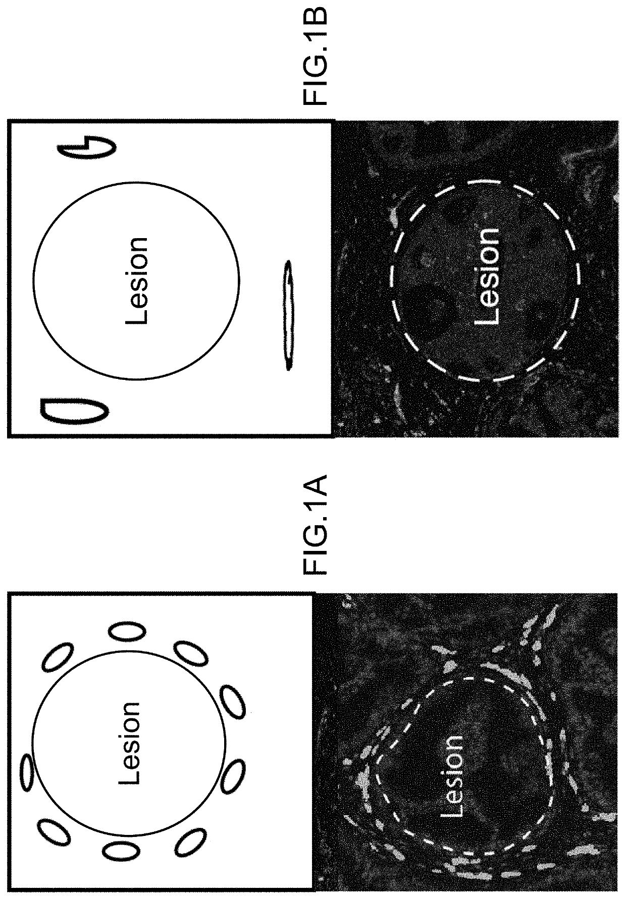
[0084]Ductal carcinoma in situ (DCIS) is a heterogeneous group of non-invasive breast lesions commonly detected by screening mammography. A small proportion of women diagnosed with DCIS subsequently develop potentially lethal invasive breast cancer (IBC). Ductal carcinoma in situ (DCIS) is the accumulation of neoplastic cells within the ducto-lobular system of the breast. Lack of invasion into the adjacent stroma is the key histological feature differentiating DCIS from invasive breast cancer (IBC). To date, there is currently insufficient data on the clonal relationship between primary DCIS and subsequent IBC to estimate the proportion of DCIS lesions actually capable of progressing to IBC, if left untreated.

[0085]DCIS currently accounts for 25-30% of all breast cancer diagnoses (60,000-70,000 cases per year in USA). DCIS is nearly universally treated by mastectomy or lumpectomy follow...

example 2

culature Abundance in Colon

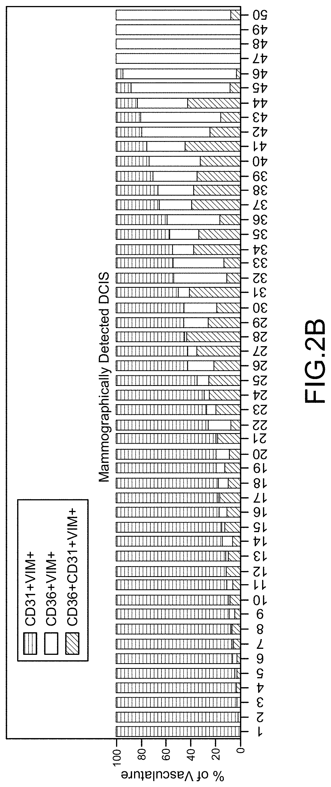
[0102]The abundance of CD36-expressing vasculature was measured in stromal tissue adjacent to healthy colon epithelia (5 subject), in stromal tissue adjacent to IBD lesions (9 subjects), and in stromal tissue adjacent to invasive colon cancer tumors (8 subjects). The abundance of CD36-expressing vasculature (CD36+ / CD31−+CD36+ / CD31+) was measured as the percentage of all vasculature [CD36+ / CD31−+CD36+ / CD31++CD36− / CD31+]. The percentage of CD36-expressing vasculature was high in healthy tissues (˜70-95%) and substantially lower in stromal tissue adjacent to invasive colon tumors (˜30% or lower in seven of eight subjects). The abundance of CD36-expressing vasculature varied in stromal tissues surrounding IBD lesions. These results suggest repression of CD36-expressing vasculature in the progression from healthy to cancerous tissue.


PUM

PropertyMeasurementUnit
threshold valueaaaaaaaaaa
thresholdaaaaaaaaaa

Abstract

Repression of CD36 vasculature in stromal tissues surrounding a pre-cancerous lesion provides is indicative of increased risk of subsequent progression to invasive cancer. Methods of assessing progression risk by measuring the abundance of CD36 vasculature in stromal tissues surrounding a precancerous lesion are provided, useful for various cancer types, including for assessing the risk of DCIS progression to breast cancer. Immunohistochemical analysis methods and diagnostic thresholds are provided, as well as associated methods of treatment and diagnostic kits.

Description

CROSS-RELATED APPLICATIONS[0001]This application is a 35 USC § 371 national stage application of International Patent Application Serial Number PCT / US2020 / 013938, entitled “Cancer Progression Risk Assessment by Microvascular Phenotype,” filed Jan. 16, 2020, which claims the benefit of priority to U.S. Provisional Application Ser. No. 62 / 793,227, entitled “Cancer Prediction by Microvascular Phenotype,” filed Jan. 16, 2019 the contents of which are hereby incorporated by reference.STATEMENT REGARDING FEDERALLY SPONSORED RESEARCH OR DEVELOPMENT[0002]This invention was made with government support under grant nos. P01 CA107584, P50 CA058207, R01 CA163687, R01 CA187800, and R01 CA197977 awarded by the National Institutes of Health. The government has certain rights in the invention.BACKGROUND OF THE INVENTION[0003]Clinically, the occurrence of precancerous lesions in a subject presents a treatment dilemma. Although at significantly higher risk than subjects not diagnosed with a precancer...

Claims


Application Information

Patent Timeline
Patent Type & Authority Applications(United States)
IPC IPC(8): G01N33/574G01N33/92
CPCG01N33/57415G01N2333/70596G01N33/92G01N2800/52
Inventor TLSTY, THEACARUSO, JOSEPHKERLIKOWSKE, KARLAMOLINARO, ANNETTEDEFILIPPIS, ROSAANNA
Owner RGT UNIV OF CALIFORNIA
Why Eureka
Try Eureka