Engineered Exosomes to Detect and Deplete Pro-Tumorigenic Macrophages
a technology of protumorigenic macrophages and exosomes, which is applied in the field of engineered exosomes for detecting and depleting protumorigenic macrophages, can solve the problems of few successes of solid tumors and poor survival, and achieve the effect of reducing tumor burden and tumor burden
- Summary
- Abstract
- Description
- Claims
- Application Information
AI Technical Summary
Benefits of technology
Problems solved by technology
Method used
Image
Examples
example 1
tion of Specificity of Precision Peptide In Vivo and Generation of CD206-Positive M2 Macrophage-Specific Exosomes
[0167]Methods and Materials
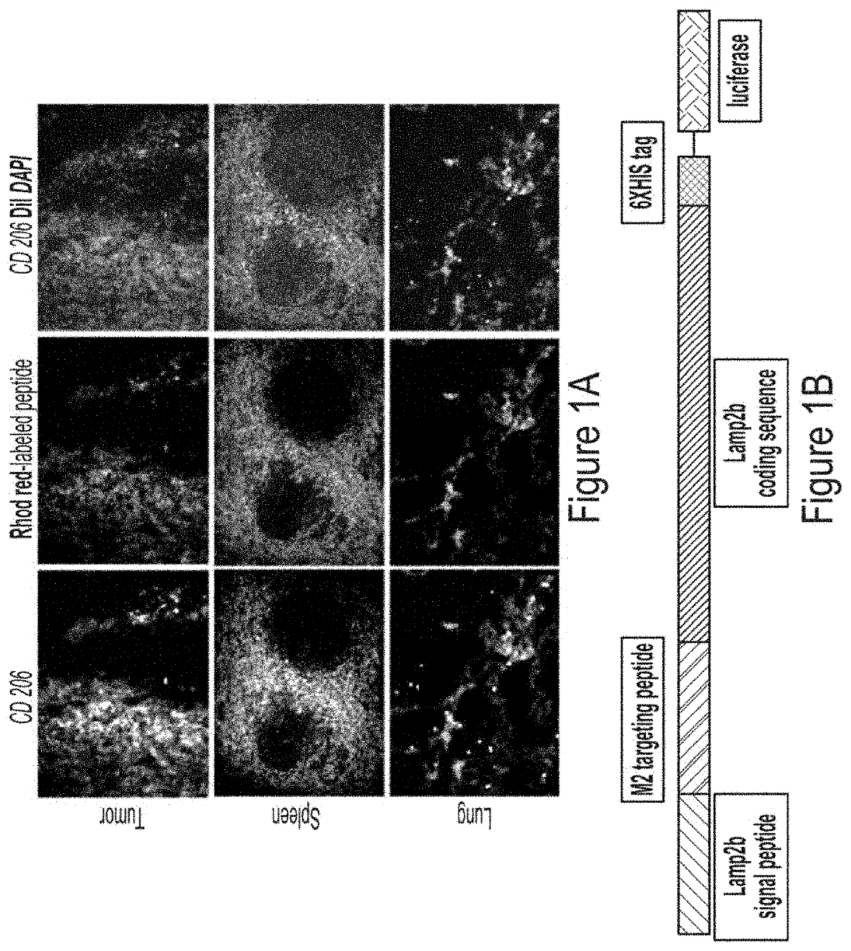
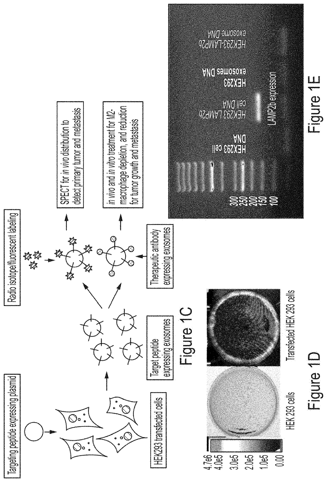
[0168]To assess in vivo targeting potential, rhodamine-labeled precision peptide (red) was injected intravenously (IV) in metastatic syngeneic murine breast cancer (4T1) bearing Balb / C mice. Three hours after injection, all animals were euthanized, and lungs, spleen and tumors were collected for immune-histochemical analysis. Frozen sections from the collected tissues were stained for CD206 (fluorescein, FITC) and counter stained with DAPI. To confer targeting potentiality, precision peptide for CD206-positive TAMs was fused to the extra-exosomal N-terminus of murine Lamp2b.
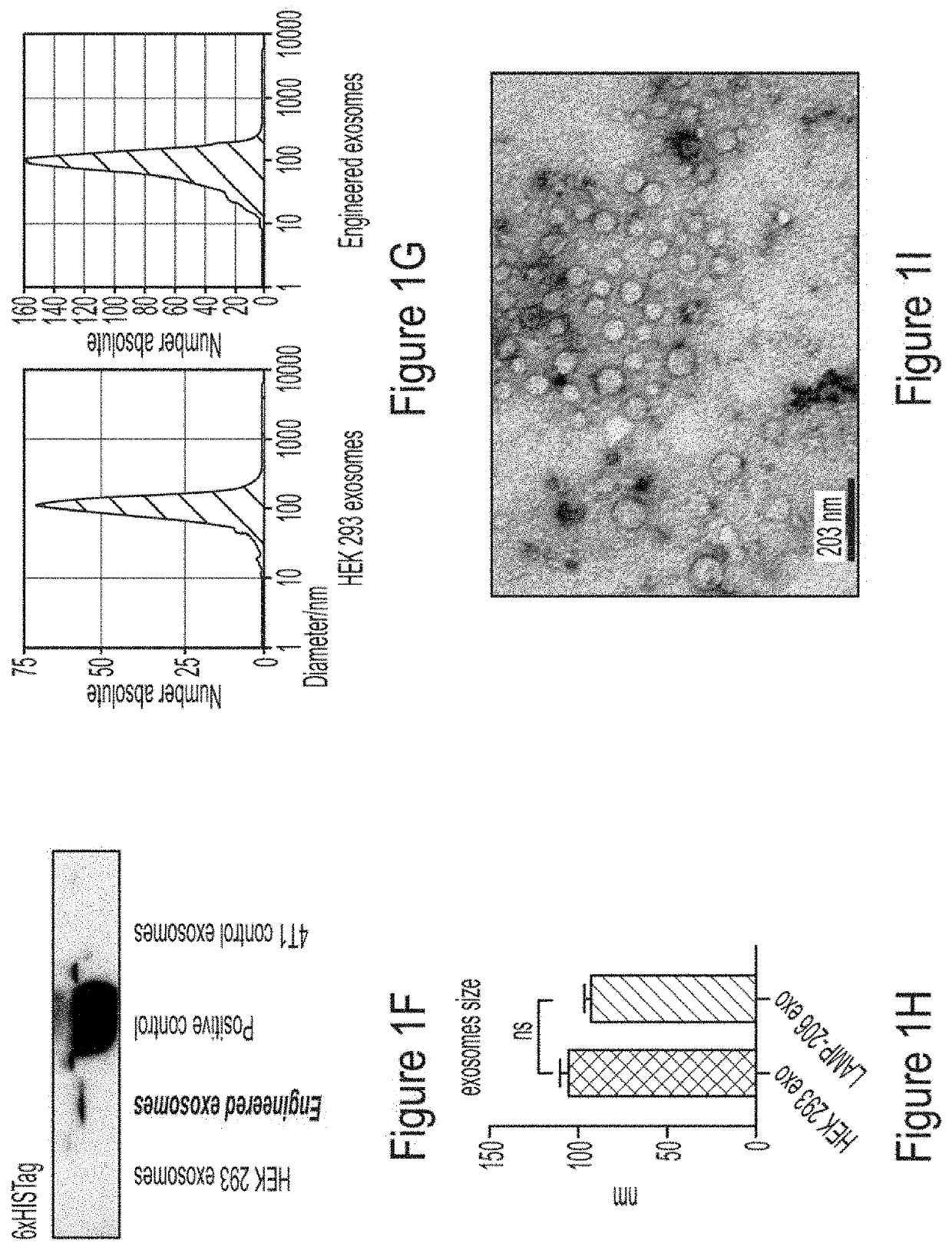
[0169]Results FIG. 1 represents generation of engineered exosomes expressing CD206-positive M2 macrophage-specific peptide along with Lamp2b. FIG. 1a exhibits immunofluorescence staining of tumor, spleen and lungs sections from 4T1 tumor-bearing mice showing co-localization o...
example 2
Potential of CD206-Positive M2-Macrophage-Specific Exosomes
[0170]Methods and Materials
[0171]To assess targeting ability of the engineered exosomes, mouse RAW264.7 macrophages towards M2-macrophages was differentiated by treating them with IL-4 and IL-3 in vitro. The cells were co-cultured with DiI-labeled (red) engineered exosomes for 4 hours followed by immunofluorescence staining for CD206-positive cells (FITC) and DAPI for nuclei.
[0172]Results
[0173]FIG. 2 represents targeting efficiency and specificity of CD206-positive M2 macrophage-specific exosomes. FIG. 2a exhibits immunofluorescence staining showing targeting potential of DiI-labeled (red) engineered exosomes. RAW264.7 mouse macrophages were differentiated to CD206-positive (FITC) cells by treating with interleukin-4 and interleukin-13. Nuclei were visualized by DAPI staining (blue). FIG. 2b exhibits immunofluorescence staining of mouse embryonic fibroblasts (MEFs) and RAW264.7 cells treated with or without anti-CD206 peptid...
example 3
and Quantification of In Vivo Distribution of CD206-Positive M2 Macrophages Targeting Exosomes
[0174]Methods and Materials
[0175]To investigate the validity of engineered exosomes as an imaging probe to determine the distribution of M2-macrophages, FDA approved clinically relevant SPECT scanning and labeling with 111In-oxine was used according to our previous study (Arbab et al., BMC Med. Imaging 2012, 12, 33). 111In-oxine-labeled non-engineered control exosomes (HEK293 exo) in metastatic (4T1) mouse breast cancer models, and engineered exosomes (M2-targeting exo) expressing precision peptide treated with either vehicle or clodronate liposome (Clophosome®-A) 24 hours before the IV administration of 111In-oxine-labeled exosomes and SPECT studies was used. Clophosome®-A is composed of anionic lipids and depletes more than 90% macrophages in spleen after a single intravenous injection (Li et al., Scient. Rep. 2016, 6, 22143-22143; Kobayashi et al., J. Biol. Chem. 2015, 290, 12603-12613)....
PUM
 Abstract
Description
Claims
Application Information
 - R&D
- Intellectual Property
- Life Sciences
- Materials
- Tech Scout
- Unparalleled Data Quality
- Higher Quality Content
- 60% Fewer Hallucinations
Browse by: Latest US Patents, China's latest patents, Technical Efficacy Thesaurus, Application Domain, Technology Topic, Popular Technical Reports.
© 2025 PatSnap. All rights reserved.Legal|Privacy policy|Modern Slavery Act Transparency Statement|Sitemap|About US| Contact US: help@patsnap.com


