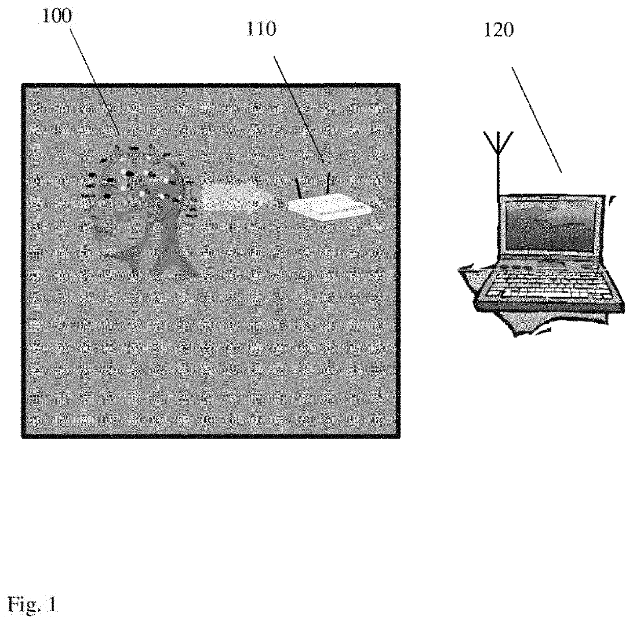
Method and system for mapping brain dysfunction for psychiatric and neurological disorders
a brain dysfunction and brain technology, applied in the field of brain mapping, can solve the problems of disability or even death, limited clinical application of topographic electroencephalography (eeg) mapping methods, and all the above techniques have serious problems
- Summary
- Abstract
- Description
- Claims
- Application Information
AI Technical Summary
Benefits of technology
Problems solved by technology
Method used
Image

Examples
example 1
of Injured Networks in Parkinson with Network Fragmentation Maps
[0061]Network fragmentation quantifies the randomness of brain interactions estimated based on dynamic cross-entropy (DCE) values. Network injuries are located in brain regions with high, abnormal values of network fragmentation. Resting state EEG was recorded using a 32-electrode cap with Ag / AgCl electrodes and then amplified. The bandwidth of the amplifiers was between (0.016-500 Hz) and data was sampled at 1000 Hz. An additional 250 Hz low-pass band filter was used and the impedance at all recording electrodes was less than 5 kΩ. Horizontal eye movements and vertical eye movements were recorded with electrodes placed near the outer cantus of each eye respectively above and below the center of the left bottom eyelid. The common average reference is used to decrease the confounding effect of brain activity.
[0062]For each data set the raw EFG resting state data is split into segments of 1-second duration and segments th...
example 2
of Injured Networks in Parkinson with Network Fragmentation Maps
[0064]In another embodiment of the invention, the regions with increased network fragmentation can be separated. This novel method is used to identify the injured brain circuitry and presented in transparent views.
[0065]For the selected patient with Parkinson and severe motor disability FIG. 12 displays sagittal transparent view that shows the presence of injured brain circuitry in centro-parietal regions 1220 and subcortical regions that include thalamic nuclei 1210. Frontal transparent view shows the presence of injured brain circuitry in centro-parietal regions 1240 and subcortical regions that include thalamic nuclei 1230.
[0066]Once the location of injured brain network is known, either non-invasive stimulation or various drugs can provide therapy. The method can also be used to monitor the effects of therapy and measure the effect of therapy on injured networks, or if the drug has targeted the required region. Drug...
PUM
 Abstract
Description
Claims
Application Information
 - R&D
- Intellectual Property
- Life Sciences
- Materials
- Tech Scout
- Unparalleled Data Quality
- Higher Quality Content
- 60% Fewer Hallucinations
Browse by: Latest US Patents, China's latest patents, Technical Efficacy Thesaurus, Application Domain, Technology Topic, Popular Technical Reports.
© 2025 PatSnap. All rights reserved.Legal|Privacy policy|Modern Slavery Act Transparency Statement|Sitemap|About US| Contact US: help@patsnap.com