
A miniature microscope for multi-contrast optical imaging in animals

a multi-contrast optical imaging and miniature microscope technology, applied in the field of miniature microscopes for multi-contrast optical imaging in animals, can solve the problems of limiting the number of contrast mechanisms that can be realized in one system, bulky hardware and optical components of the custom-built imaging system, and the inability to carry most multi-contrast neuroimaging systems

Pending Publication Date: 2020-07-16
THE JOHN HOPKINS UNIV SCHOOL OF MEDICINE
View PDF0 Cites 1 Cited by
  • Summary
  • Abstract
  • Description
  • Claims
  • Application Information

AI Technical Summary

Benefits of technology

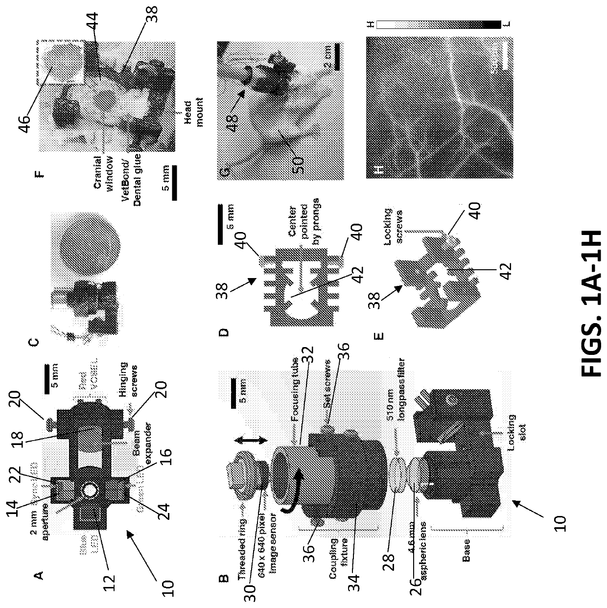
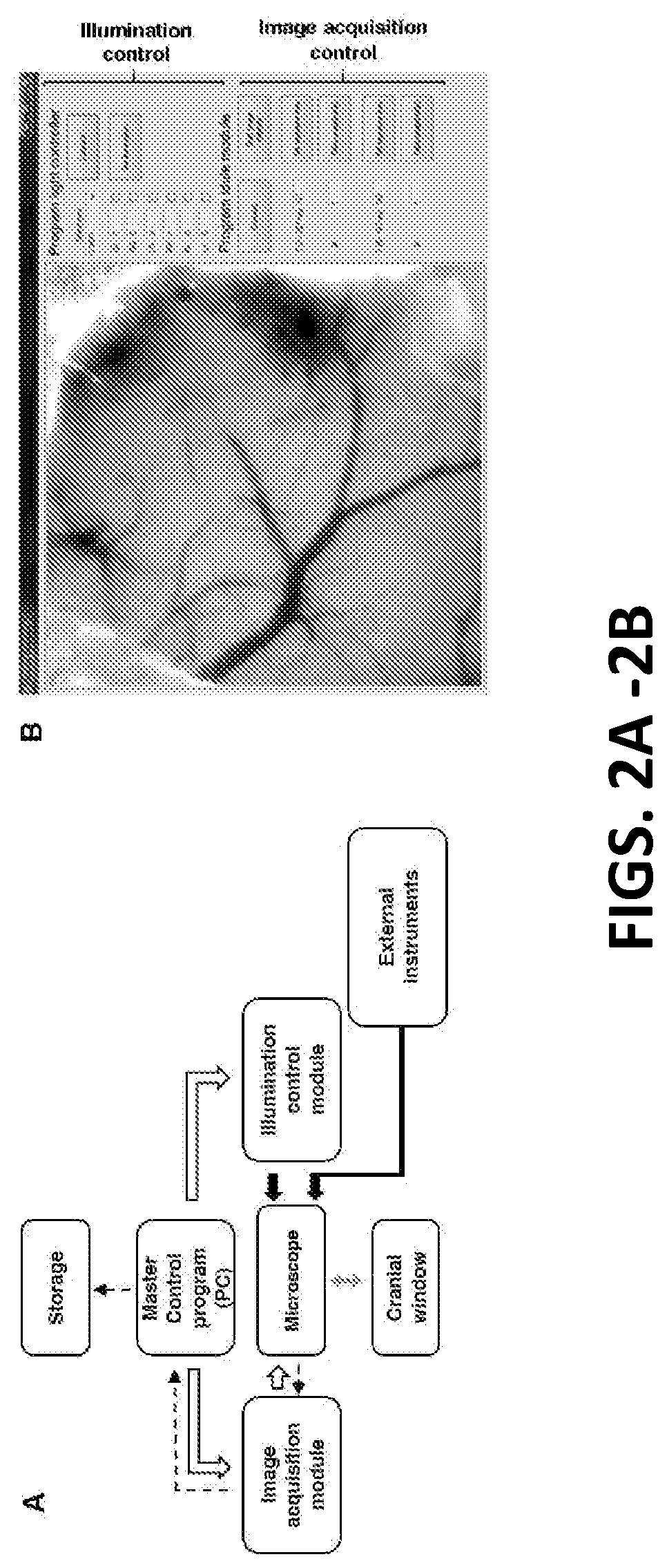
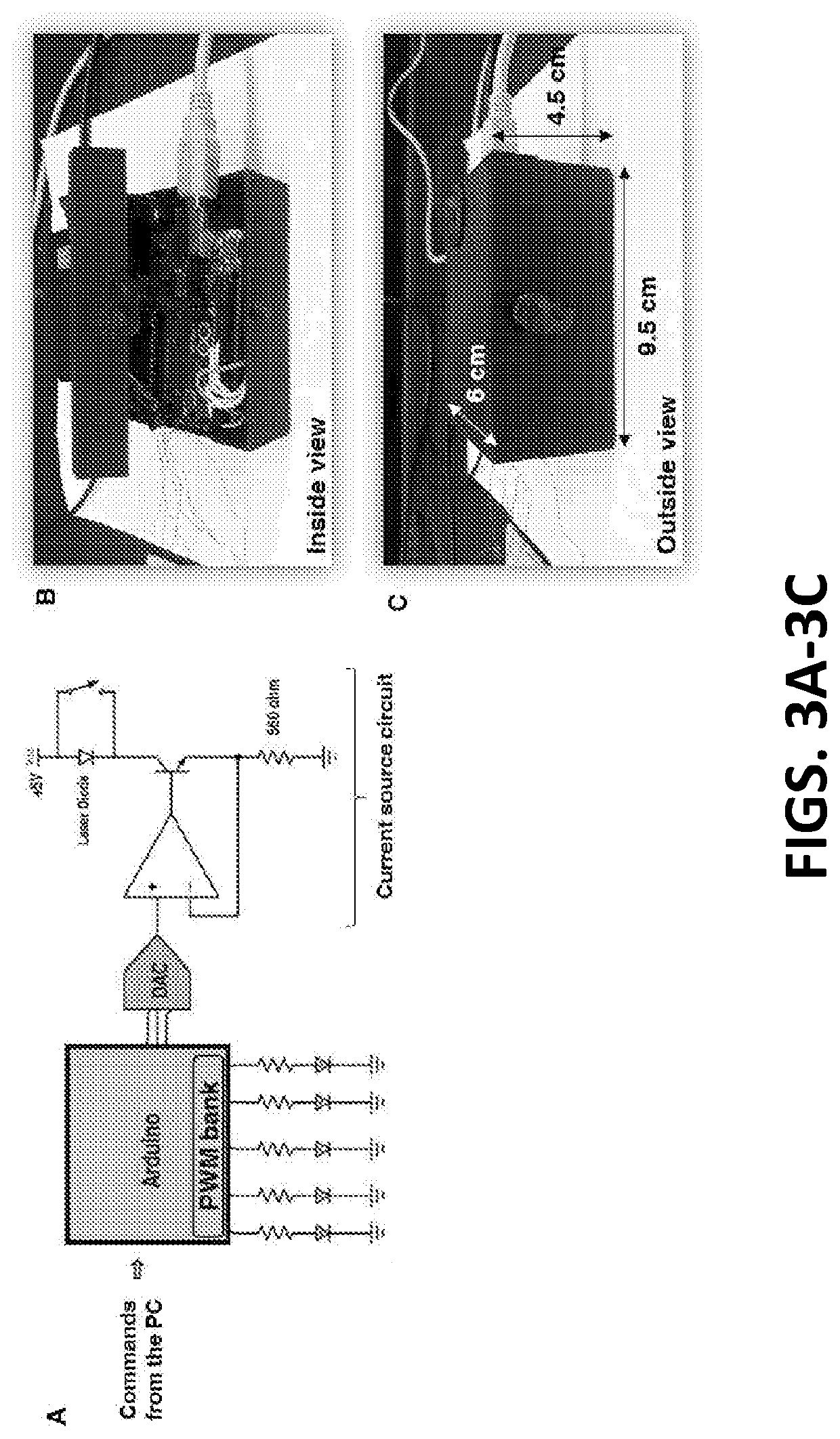
The present invention is a device for wide-field optical imaging that includes a microscope for multi-contrast imaging. The microscope has an optical filter for cutting off blue fluorescent excitation light and can be mounted on different body locations of a subject animal. The microscope includes custom-built mounts for variable magnification imaging and a wireless module for tether-free operation. It also includes integrated circuits or very-large-scale integration fabricated chips with an on-board image sensor and modules for hardware encoded image processing and wireless transmission. The microscope also has a remote master controller for controlling imaging and illumination parameters, a data transmission system, and an illumination controller for controlling the illumination sources. The wireless connection takes the form of WiFi or Bluetooth®. The technical effect of this invention is to provide a versatile and efficient tool for wide-field optical imaging in animal research.

Problems solved by technology

The need to combine multiple optical contrast mechanisms often requires a custom-built imaging system with bulky hardware and optical components.
This makes most multi-contrast neuroimaging systems non-portable, precluding their use in a wide variety of in vivo experiments in freely moving or unanesthetized animals.
Finally, the bulkiness of conventional multi-contrast imaging systems limits the number of contrast mechanisms that can be realized in one system, and also restricts the recording of additional neurophysiological variables such as electrophysiology and neurochemical recordings.
However, currently available imaging systems are limited to performing eithef neural or hemodynamic imaging, but not both.
This in turn has limited in viva experiments in awake animals to those that interrogate either the brain's neuronal or vascular function, but not both.
However, they lack the ability to directly interrogate neural function, which requires the ability to image fluorescence (FL).
However, due to technical limitations, current miniature microscopes that have a FL channel do not perform IOS and LSC imaging.

Method used


Image

  • A miniature microscope for multi-contrast optical imaging in animals
  • A miniature microscope for multi-contrast optical imaging in animals
  • A miniature microscope for multi-contrast optical imaging in animals

Examples


Embodiment Construction

[0025]The presently disclosed subject matter now will be described more fully hereinafter with reference to the accompanying Drawings, in which some, but not all embodiments of the inventions are shown. Like numbers refer to like elements throughout.

[0026]The presently disclosed subject matter may be embodied in many different forms and should not be construed as limited to the embodiments set forth herein; rather, these embodiments are provided so that this disclosure will satisfy applicable legal requirements. Indeed, many modifications and other embodiments of the presently disclosed subject matter set forth herein will come to mind to one skilled in the art to which the presently disclosed subject matter pertains having the benefit of the teachings presented in the foregoing descriptions and the associated Drawings. Therefore, it is to be understood that the presently disclosed subject matter is not to be limited to the specific embodiments disclosed and that modifications and o...


PUM


Abstract

An embodiment in accordance with the present invention provides a miniature microscope capable of performing in vivo, real-time imaging of multiple organ sites in awake and behaving animals (e.g. rodents). A microscope according to the p resent invention includes multiple optical contrast mechanisms (i.e. contrast arising from neural, hemodynamic and other physiological components). Exemplary contrast mechanisms include, but are not limited to fluorescence, hemoglobin level, deoxyhemoglobin level, and blood flow. The microscope is fully adaptable to in vitro and ex vivo imaging, can be customized to concurrently image at variable magnifications, conduct optogenetic / electrical / chemical stimulations, drug delivery, micro-dialysis, accompanied by electrical signal recording, wireless image transmission and charging.

Description

CROSS REFERENCE TO RELATED APPLICATIONS[0001]This application claims the benefit of U.S. Provisional Patent Application No. 62 / 529,100 filed Jul. 6, 2017, which is incorporated by reference herein, in its entirety.GOVERNMENT SUPPORT[0002]This invention was made with government support under grant number 1R21CA175784-01 awarded by the National Institutes of Health. The government has certain rights in the invention.FIELD OF THE INVENTION[0003]The present invention relates generally to imaging devices. More particularly, the present invention relates to a miniature microscope for multi-contrast (e.g. contrast arising from neural, hemodynamic or other physiologic components) optical imaging in animals or in vitro preparations such as cellular or perfusion assays.BACKGROUND OF THE INVENTION[0004]Brain function involves the interplay between many biological variables (e.g. a complex set of interactions between neurons, blood vessels, astrocytes etc.). Therefore, to better understand brai...

Claims


Application Information

Patent Timeline
Patent Type & Authority Applications(United States)
IPC IPC(8): G02B21/16A61B5/00A61B5/1455G02B21/06G02B21/02G02B21/36H05B47/19
CPCG02B21/365A61B5/0071A61B5/002A61B5/0042A61B5/1455H05B47/19G02B21/025G02B21/06A61B5/6814G02B21/16A61B5/0077A61B1/00A61B5/0075A61B5/0084A61B5/0261A61B5/489G02B21/0008G02B21/082
Inventor PATHAK, ARVINDTHAKOR, NITISHSENARATHNA, JANAKAYU, HANG
Owner THE JOHN HOPKINS UNIV SCHOOL OF MEDICINE
Learn More