
Regenerating functional neurons for treatment of neural injury caused by disruption of blood flow

a functional neuron and neural injury technology, applied in the direction of peptides, drug compositions, peptides, etc., can solve the problems of pathological changes in the vicinity of blood flow interruption, often killed or injured neurons and other cns cells, etc., to inhibit axonal regeneration and neural regrowth, improve the effect of defense response, and worsen the infarct area

Pending Publication Date: 2020-02-20
PENN STATE RES FOUND
View PDF2 Cites 1 Cited by
  • Summary
  • Abstract
  • Description
  • Claims
  • Application Information

AI Technical Summary

Benefits of technology

The present invention provides a method for treating neurological disorders by using in vivo cell conversion technology to directly convert reactive glial cells into functional neurons. This technology has advantages over traditional stem cell therapy, including the use of endogenous glial cells and reduced potential tumor risk. Additionally, the technology can regenerate a large number of new neurons in areas of disruption of normal blood flow, such as stroke areas, and can also reduce the number of reactive astrocytes and the inflammatory factors secreted by them. Overall, this technology has shown promising results in restoring motor functions and promoting brain recovery after ischemic stroke.

Problems solved by technology

Neurons and other CNS cells are often killed or injured as a result of a disease or injury which interrupts blood flow in the region where the cells are located.
Furthermore, pathological changes in the vicinity of the blood flow interruption are responsible for numerous adverse effects, such as destruction or injury of neurons, destruction or injury of glial cells, destruction or injury of blood vessels, and destruction or injury of supporting cells, in the region of the CNS affected by the disruption of normal blood flow.

Method used


Image

  • Regenerating functional neurons for treatment of neural injury caused by disruption of blood flow
  • Regenerating functional neurons for treatment of neural injury caused by disruption of blood flow
  • Regenerating functional neurons for treatment of neural injury caused by disruption of blood flow

Examples


example 1

onverts Stroke-Induced Reactive Astrocytes into Neurons

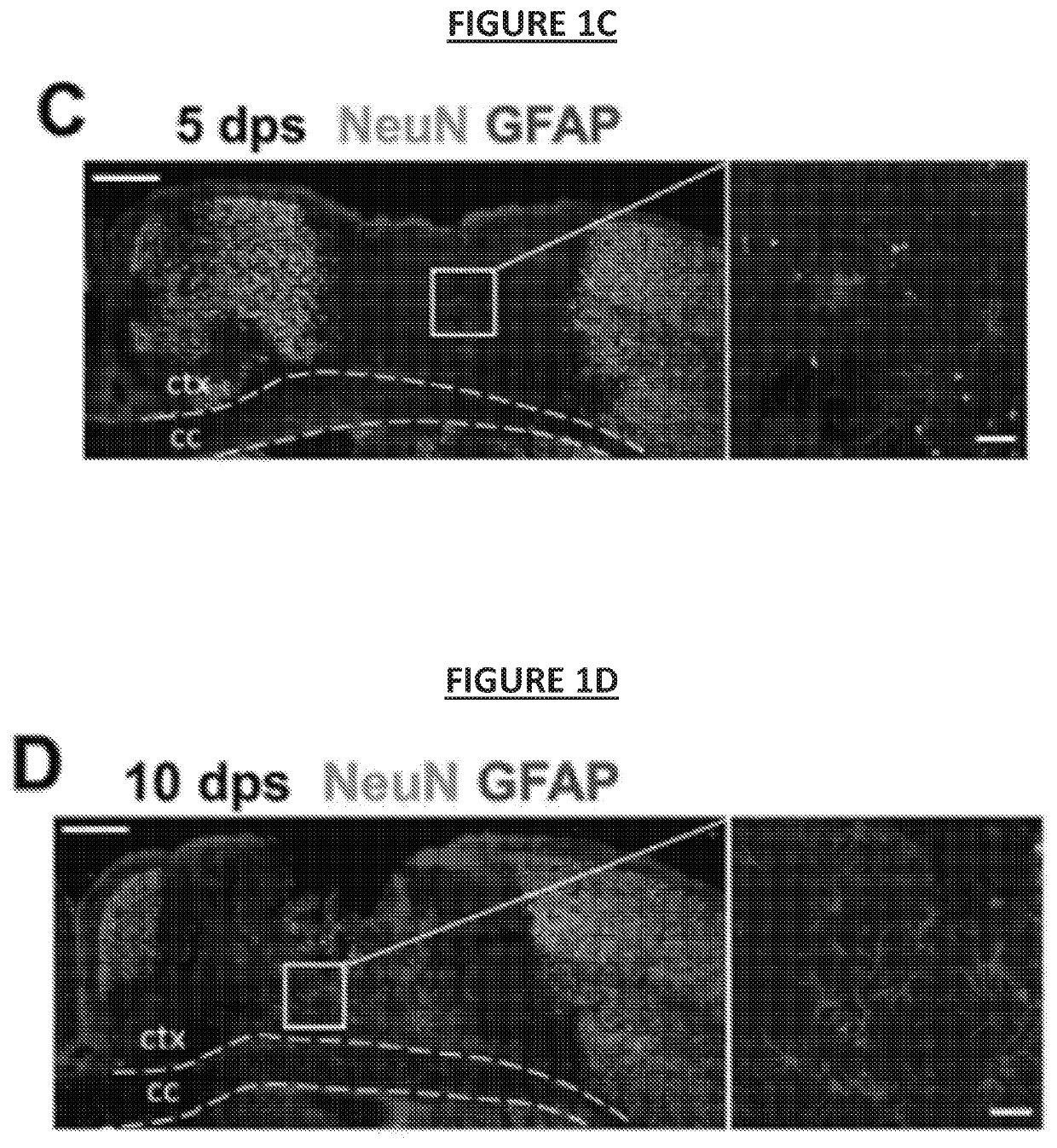
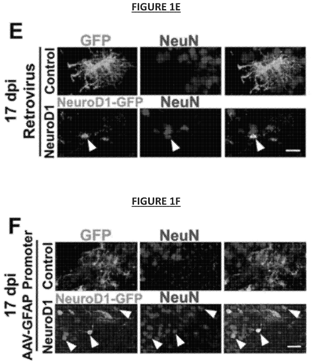
[0303]In this example, it was investigated whether in vivo cell conversion can achieve functional brain repair following an ischemic stroke. A focal stroke model induced by the vasoconstrictive peptide endothelin-1 (ET-1) was used to produce consistent local ischemic injury in rodents (Fuxe et al., 1997; Hughes et al., 2003; Roome et al., 2014; Windle et al., 2006).

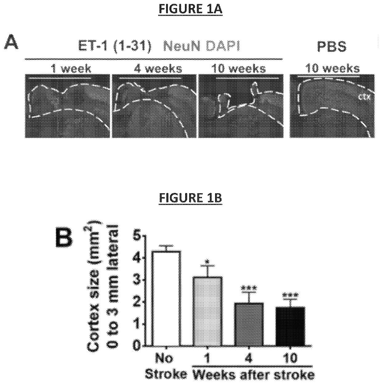
[0304]When two different ET-1 peptides, one with 21 amino acids (ET-1 (1-21)) and another with 31 amino acids (ET-1 (1-31)) were compared, it was discovered that ET-1 (1-31) produced more severe and prolonged brain damage than ET-1 (1-21) (see, FIG. 2A). A genetic strain of mice with FVB background gave more severe stroke damage than the commonly used B6 / C57 mice (see, FIG. 2A). Notably, significant cortical tissue loss was observed with injection of ET-1 (1-31) into the motor cortex of FVB mice (see, FIG. 1A and FIG. 1B)—establishing a severe focal stroke model with ...

example 2

of Neuroinflammation After Cell Conversion

[0317]Accompanying the astrocyte-to-neuron conversion after NeuroD1 infection in the stroke areas, a significant reduction in GFAP signal in the stroke areas was observed (see, FIG. 5A). This is expected, because >70% of NeuroD1-infected reactive astrocytes have been converted into neurons, and the GFAP signal will be decreased accordingly. However, this also raises a question whether astrocytes might be depleted in the converted areas. To answer this, when the astrocyte-to-neuron conversion was largely completed, the stroke areas close to the infarction (*) which were highly infected by NeuroD1 AAV at 17 days post viral injection (dpi; 27 dps) were examined.

[0318]While astrocytes did show a reduction in the NeuroD1-infected areas, there were still a significant number of astrocytes remaining (see, FIG. 5B). Surprisingly, not only was the astrocyte number reduced, but astrocyte morphology was significantly changed in the NeuroD1-converted ar...

example 3

neration Leads to Neuroprotection

[0325]Consistent with a reduction of neuroinflammation, the number of NeuN-positive neurons appeared to increase significantly in the NeuroD1-infected areas (see, FIG. 7A). Surprisingly, in the stroke areas close to the injury core, not only were NeuroD1-GFP-labeled neurons detected but also many non-converted neurons that were neither GFP-positive (see, FIG. 7A, arrow) nor NeuroD1-positive (see, FIG. 7B, arrow).

[0326]Quantitative analysis revealed that ˜40% of NeuN+ neurons in the peri-infarct areas were NeuroD1-positive and ˜60% of neurons were NeuroD 1-negative (see, FIG. 7C; Control group NeuN+, 27.1±8.1 / 0.1 mm2; NeuroD1 group: NeuroD1− / NeuN+, 64.9,±7.9 / 0.1 mm2; NeuroD1+ / NeuN+, 39.9,±2.4 / 0.1 mm2; n=3 mice per group).

[0327]Similar results were obtained when the GFP+ / NeuN+ neurons in NeuroD1-NeuroD1-GFP infected neurons were quantified (see, FIG. 8A). In addition, the non-converted neurons in the NeuroD1-treated areas also more than doubled the num...


PUM

PropertyMeasurementUnit
Volumeaaaaaaaaaa
Volumetric flow rateaaaaaaaaaa
Flow rateaaaaaaaaaa

Abstract

Methods of treating the effects of disruption of normal blood flow in the CNS in an individual subject in need thereof are provided according to aspects of the present invention which include administering a therapeutically effective dose of exogenous NeuroD1 to an area where normal blood flow has been disrupted. Compositions are provided including 1) a recombinant adeno-associated adenovirus expression vector comprising a glial cell specific promoter operably linked to a nucleic acid encoding a site-specific recombinase and 2) a recombinant adeno-associated adenovirus expression vector comprising a ubiquitous promoter operably linked to a nucleic acid encoding NeuroD1, wherein the nucleic acid encoding NeuroD1 is inverted and flanked by two sets of site-specific recombinase recognition sites such that action of the recombinase irreversibly inverts the nucleic acid encoding NeuroD1 such that NeuroD1 is expressed in a mammalian cell.

Description

REFERENCE TO RELATED APPLICATIONS[0001]This application claims priority to U.S. Provisional Patent Application Ser. No. 62 / 464,469, filed Feb. 28, 2017; and U.S. Provisional Patent Application Ser. No. 62 / 518,914, filed Jun. 13, 2017. The entire content of each application is incorporated herein by reference.GOVERNMENT SUPPORT[0002]This invention was made with government support under Grant No. AG045656, awarded by the National Institutes of Health. The Government has certain rights in the invention.FIELD OF THE INVENTION[0003]This invention relates to compositions and methods for treating the effects of disruption of normal blood flow in the CNS of an individual subject.BACKGROUND OF THE INVENTION[0004]The central nervous system (CNS) in mammals is largely unable to regenerate itself following injury. Neurons and other CNS cells are often killed or injured as a result of a disease or injury which interrupts blood flow in the region where the cells are located. Furthermore, patholog...

Claims


Application Information

Patent Timeline
IPC IPC(8): A61K38/17C12N15/86A61P25/00
CPCC12N2830/008A61K38/1709A61K48/00C12N2750/14132C12N2750/14143C12N2800/30C12N15/86A61P25/00A61K48/0058C12N2800/40C07K14/4702A61K38/00
Inventor CHEN, GONG
Owner PENN STATE RES FOUND
Learn More