
Systems and methods for improving placement of devices for neural stimulation

a neural stimulation and placement system technology, applied in the field of systems and methods for improving the placement of neural stimulation devices, can solve problems such as noise recording, sub-optimal or ineffective stimulation of neural tissue, and inaccuracy in decoding

Pending Publication Date: 2020-01-16
SYNCHRON AUSTRALIA PTY LTD
View PDF8 Cites 5 Cited by
  • Summary
  • Abstract
  • Description
  • Claims
  • Application Information

AI Technical Summary

Benefits of technology

The patent text describes a system and method for implanting electrodes in blood vessels in the brain and other regions of the body. The system includes an activity image that shows neural activity in the region, a structural image that shows the anatomical structure of the vascular network, and a real-time image that shows the region in real-time. The activity image is co-registered with the structural image to identify a target location within the vascular network. The system can also include a virtual marker that is displayed on the real-time image to provide guidance for implanting the medical device. The technical effect of this system is to enable accurate and safe implantation of medical devices in specific locations within the body, particularly in regions with complex anatomy such as the brain.

Problems solved by technology

Sub-optimal placement of recording devices (e.g., electrode arrays) can lead to recording of noise, and thus decoding inaccuracies.
Sub-optimal placement of stimulating devices (e.g., electrode arrays) can also lead to sub-optimal or ineffective stimulation of neural tissue.
Neuroendovascular surgeries are often radiologically guided, for example, with intra-operative magnetic resonance imaging as well as with intra-operative computed tomography (CT), where the interventionist can view the outline of the vessels, but such surgical techniques do not currently provide the interventionist with static or real-time functional and / or physical reference marks for determining whether a current position of a recording and / or stimulating device (e.g., an electrode array) within a vessel is near the preferred target cortical location prior to, during, or after deployment of the device.
Physical references alone such as vessel tortuosity, vessel size, aneurism location and blockage locations currently do not allow interventionists to determine whether the position of the device is in the preferred target cortical location, as such physical references are currently not associated with or linked to any functional activity.
Further, whether the cortical location adjacent to the current device position inside the vessel is the desired cortical location for thought-decoding cannot currently be determined from within the vessel, as the cortex is not exposed at all during the minimally invasive endovascular surgery and cannot be stimulated intraoperatively, which is the status quo of open-brain surgery.

Method used


Image

  • Systems and methods for improving placement of devices for neural stimulation
  • Systems and methods for improving placement of devices for neural stimulation
  • Systems and methods for improving placement of devices for neural stimulation

Examples


Embodiment Construction

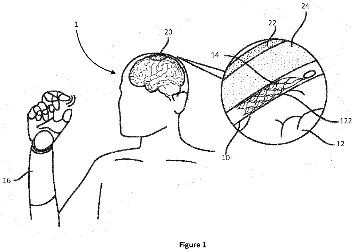
[0035]This disclosure is not limited to the particular embodiments, variations, or examples described, as such may, of course, vary. For example, while neural tissue in the brain is referred to throughout the disclosure, the disclosure is applicable to any tissue that emits signals that can be recorded and / or has cells which can be stimulated. This can include tissue and vessels anywhere in the body, for example, the brain, the neck, extremities, and torso.

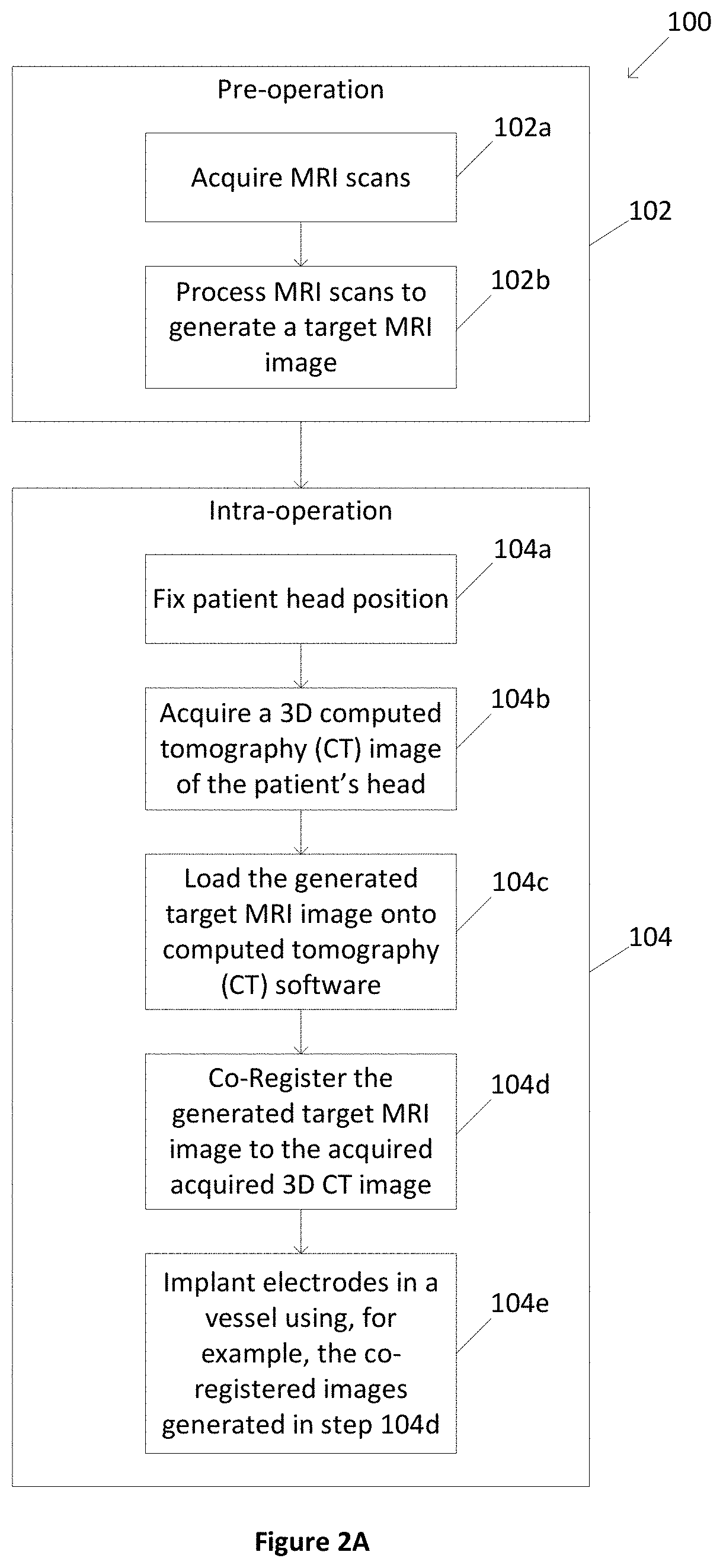
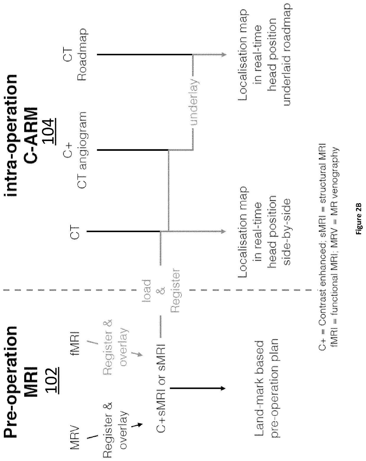
[0036]Structural and functional imaging techniques can be used to design a surgical plan, where the surgical plan can have one or multiple target locations for a device, for example, 1 to 5 or more target locations, including every 1 target location increment within this range (e.g., 1 target location, 2 target locations, 3 target locations, . . . , 5 target locations). Clearly, the number of target locations can vary depending upon the intended application. The target locations can be ranked from most preferred to least preferred...


PUM


Abstract

The present invention involves systems and methods for creating and displaying surgical plans for implanting devices in the body.

Description

RELATED APPLICATION[0001]This is a non-provisional application of U.S. Provisional application No. 62 / 696,628 filed on Jul. 11, 2018, the entirety of which is incorporated by reference.BACKGROUND[0002]Signal generating and / or receiving devices can be implanted in or on the body to stimulate tissue, record signals from tissue, or both. For example, electrodes can be implanted in or on the body to stimulate tissue, record signals from tissue, or both.[0003]Implanting electrodes in or near a desired (e.g., ideal) cortical location can influence the efficacy of stimulating and / or recording neural tissue (e.g., using brain-computer-interfaces) such as decoding thoughts from neural signals. Sub-optimal placement of recording devices (e.g., electrode arrays) can lead to recording of noise, and thus decoding inaccuracies. Sub-optimal placement of stimulating devices (e.g., electrode arrays) can also lead to sub-optimal or ineffective stimulation of neural tissue. This is especially true, fo...

Claims


Application Information

Patent Timeline
IPC IPC(8): A61N1/05A61B90/00A61B34/10A61N1/36
CPCA61N1/0534A61B34/10A61B2090/3762A61B90/37A61N1/3605A61B2090/374A61B2090/364A61N1/0531A61N1/056A61B6/037A61B6/5247A61B6/5235A61B6/501A61B6/504A61B5/0042A61B5/055A61B6/463
Inventor YOO, PETER ELI
Owner SYNCHRON AUSTRALIA PTY LTD
Learn More