
Transcatheter device for interatrial anastomosis

a transcatheter device and interatrial anastomosis technology, applied in the field of transcatheter devices for interatrial anastomosis, can solve the problems of significant associated healthcare costs and major epidemic of heart failure, and achieve the effects of increasing the size of the cutter, and slowing the natural healing process of the tissu

Pending Publication Date: 2019-12-12
TEXAS MEDICAL CENT
View PDF1 Cites 6 Cited by
  • Summary
  • Abstract
  • Description
  • Claims
  • Application Information

AI Technical Summary

Benefits of technology

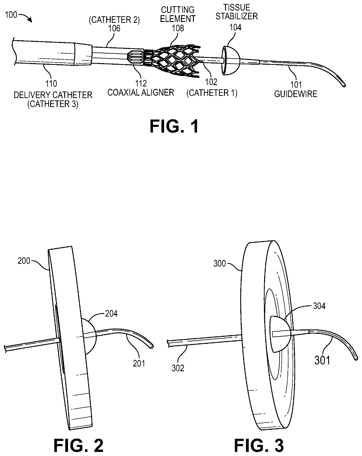
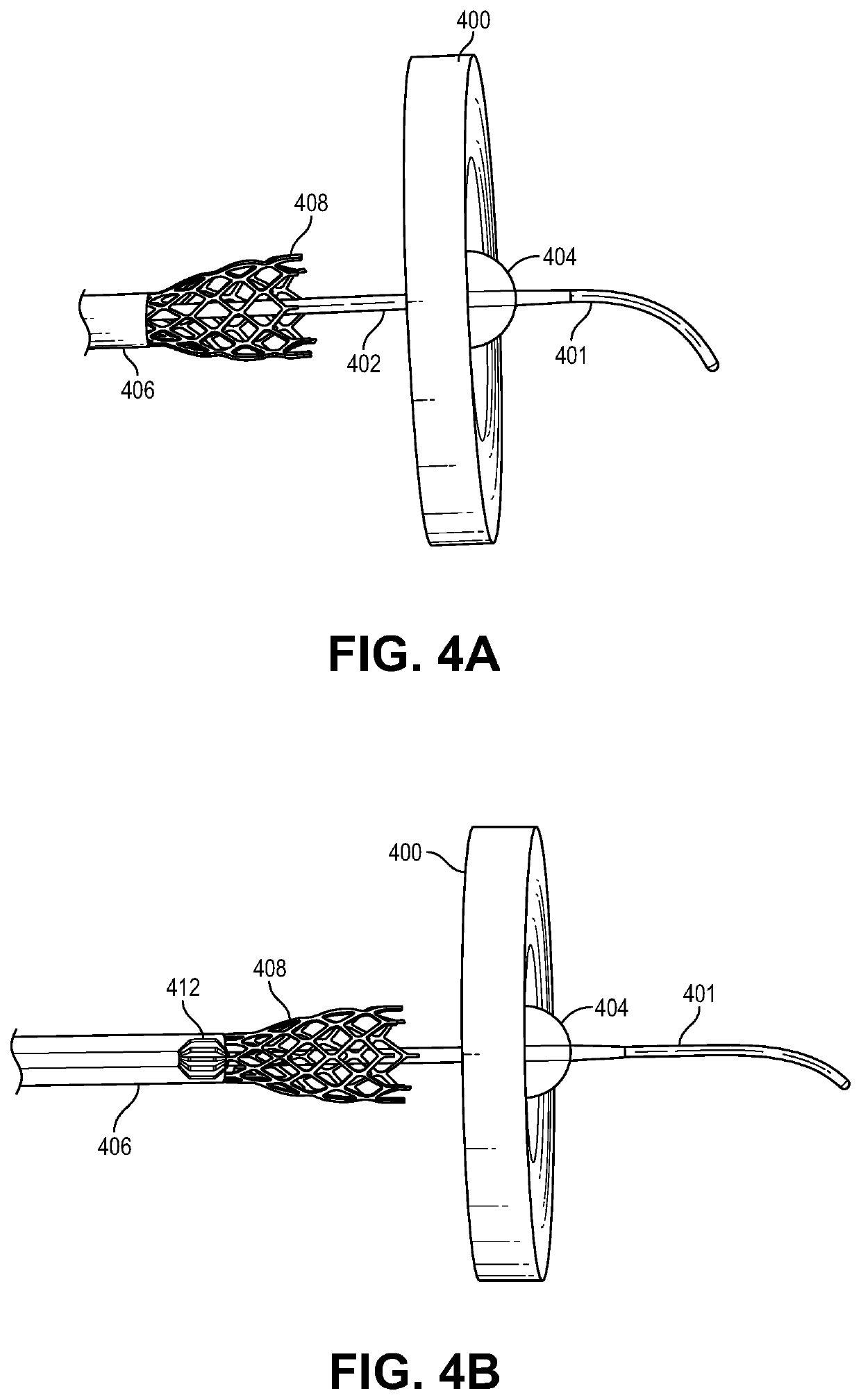
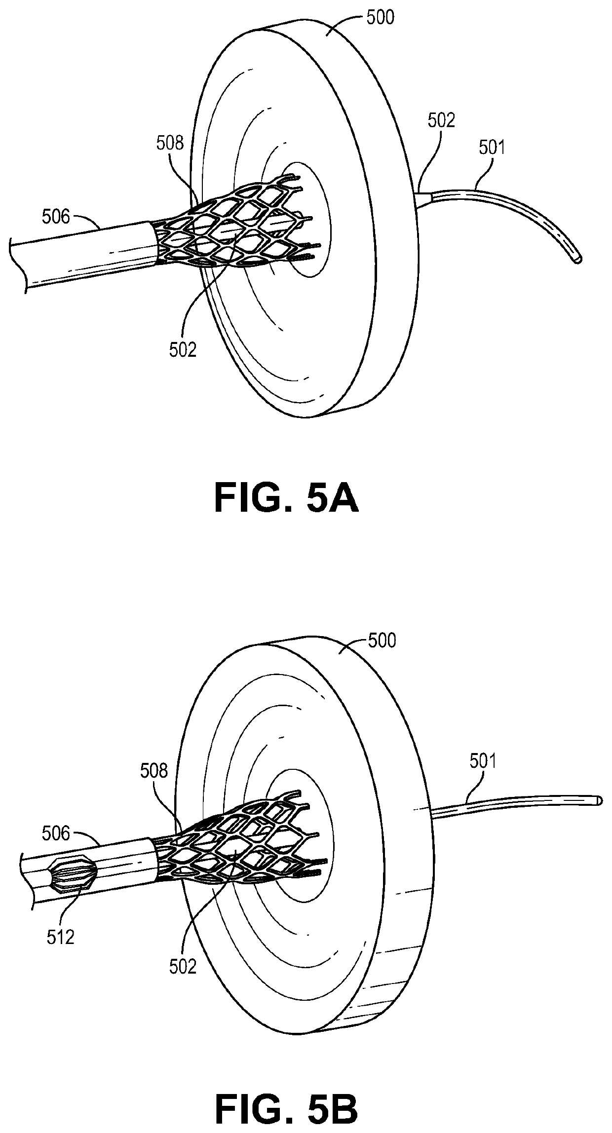
This patent describes device assemblies for treating heart failure. The device includes a delivery catheter, a first internal coaxial catheter with a stabilizer, a second internal coaxial catheter with a cutter, and a coaxial alignment mechanism. The first internal coaxial catheter has a puncture tip for crossing an interatrial septum. The device also has adjustable cutting and tissue stabilizer dimensions. The coaxial alignment mechanism provides a working track for the device assembly. The device can be used to treat heart failure by delivering a cutting tool to the interatrial septum.

Problems solved by technology

Thus, heart failure remains a major epidemic with significant associated healthcare costs.

Method used


Image

  • Transcatheter device for interatrial anastomosis
  • Transcatheter device for interatrial anastomosis
  • Transcatheter device for interatrial anastomosis

Examples


embodiment 1400

[0189]In some embodiments, as shown in FIGS. 14A-14C, the tissue stabilizer embodiment 1400 is deployed from catheter 1, 1402, tracking along a guidewire (not shown) and again takes the form of a plurality of tines 1407 that pass through the septum as they were being deployed from the catheter 1, and expand in an umbrella fashion. The tines partially pierce through the septum 1420 (with or without barbs at the end of each tine) to prevent the septum from moving off of the tines. In either embodiment (with or without barbs on the tines), after the cutter 1408 is deployed from the delivery catheter 3 1410, and a portion of the interatrial septum is excised, the expanded tines permit resheathing within the delivery catheter, over the re-collapsed tines and excised tissue 1403, followed by removal of the device assembly from the body, as described previously. In some embodiments, the process of resheathing into the delivery catheter 1410, the tines 1407 bend backwards such that the tiss...

embodiment 1500

[0190]FIGS. 15A-15E are sequential representative illustrations of an exemplary embodiment of the tissue stabilizer taking the form of a loop supported by shape-set tines. Catheter 1, 1502 comprises a cap, 1523 having a central lumen that houses the collapsed tissue stabilizer. The tissue stabilizer catheter 1524, which is slidably engaged with the outside diameter of the catheter 1, 1502, is translated until its distal tip 1501, crosses the interatrial septum and allows for unsheathing of the catheter that comprises the tissue stabilizer in the left atrium. In some embodiments, as shown in FIGS. 15A-15E, the tissue stabilizer embodiment 1500 is collapsed and housed in catheter 1, 1502, that has a penetrating tip 1501, tracking along a guidewire (not shown) and takes the form of a loop supported by shape-set tines 1504, made with a shape memory metal or alloy. In some embodiments, catheter 1, 1502 comprises a cap 1522 and a shaft 1523. FIGS. 15A-15E are sequential representative ill...


PUM


Abstract

The present disclosure relates to a device assembly and a method for treating heart failure by normalizing elevated blood pressure in the left atrium of a heart of a mammal. Disclosed herein is a transcatheter interatrial septum excision device assembly configured to create a sized interatrial aperture between the right and left atria of a heart for the relief of elevated left atrial pressure. The device assembly comprises a delivery catheter, a tissue stabilizer attached to a first catheter having a central lumen and a penetrating tip that permits passage of a guidewire, and a cutter attached to a second catheter having a central lumen that permits passage of the first catheter. Alternative configurations comprise a (third) catheter having a central lumen that permits passage of the aforementioned components to and from the right atrium, a tissue retention mechanism and an optional coaxial alignment mechanism.

Description

CROSS-REFERENCE[0001]This application is a continuation of International Patent Application No. PCT / US2018 / 017487 filed Feb. 8, 2018, which claims the benefit of U.S. Provisional Application No. 62 / 457,605, filed Feb. 10, 2017, U.S. Provisional Application No. 62 / 473,027, filed Mar. 17, 2017, U.S. Provisional Application No. 62 / 532,223, filed Jul. 13, 2017, and U.S. Provisional Application No. 62 / 558,178, filed Sep. 13, 2017, which applications are incorporated herein in their entirety by reference.BACKGROUND OF THE INVENTION[0002]Congestive heart failure (CHF) is a chronic condition affecting 6 million people in the US and 23 million people worldwide. Incidence is expected to rise in the next 10 years with 650,000 new cases diagnosed annually in the US. Once a patient is diagnosed with CHF, 5 and 10-year survival rates are estimated at 50% and 10% respectively. Heart failure is the most common cause of U.S. hospital admission in patients over 65 and accounts for almost 1 million ho...

Claims


Application Information

Patent Timeline
Patent Type & Authority Applications(United States)
IPC IPC(8): A61B17/34A61B17/3205A61M25/01A61B17/32
CPCA61B18/1492A61B2017/00867A61M25/0108A61B17/32002A61B17/3478A61B2018/0212A61B2017/22044A61B2017/3488A61B17/320068A61B2018/00357A61B2017/00247A61B17/32053A61M25/06A61B17/3209A61B17/34A61B17/11A61B18/02A61B2017/320052A61B2017/32113A61B2017/1139A61B2017/3425A61B2018/00601A61B2017/00986
Inventor AREVALOS, CHRISTOPHER ALEXANDERGREIJDANUS, ALBERTIENKRIEGEL, JACOBPATEL, AVNI
Owner TEXAS MEDICAL CENT
Learn More