
Automated phase selection for ecg-gated cardiac axial ct scans

a phase selection and axial ct scan technology, applied in the field of computed tomography, can solve the problems of distortion of the resulting tomographic image, difficulty in triggering a short scan so as to complete the short scan during a time when the heart is relatively stationary, natural variation of the heart rate, etc., and achieve the effect of reducing motion artifacts

Active Publication Date: 2019-12-12
ANLOGIC CORP (US)
View PDF0 Cites 0 Cited by
  • Summary
  • Abstract
  • Description
  • Claims
  • Application Information

AI Technical Summary

Benefits of technology

The present patent application describes a method for reducing motion artifacts in computed tomography images of anatomical objects. The method involves comparing projections and backprojections of the object acquired at different angular locations within a first range of source angles. The differences between the two are used to quantify the movement of the object during the acquisition process. Short scan data is then identified, which includes a subset of the extended scan data acquired at different locations within a second range of source angles where the object's movement is less than a certain threshold. The computed tomography image of the object is then reconstructed from the short scan data. This method helps to improve the accuracy and reliability of computed tomography images.

Problems solved by technology

However, heartrates naturally vary, even while a patient is resting.
Because the duration of time between heartbeats is not constant, triggering a short scan so the short scan can be completed during a time when the heart is relatively stationary is difficult.
The use of data acquired during the pronounced motion of the heart results in distortions to the resulting tomographic image referred to as motion artifacts.
Motion artifacts appearing in one or more of the slices to be assembled into the image of the entire heart diminish the quality of the assembled image.

Method used


Image

  • Automated phase selection for ecg-gated cardiac axial ct scans
  • Automated phase selection for ecg-gated cardiac axial ct scans
  • Automated phase selection for ecg-gated cardiac axial ct scans

Examples


Embodiment Construction

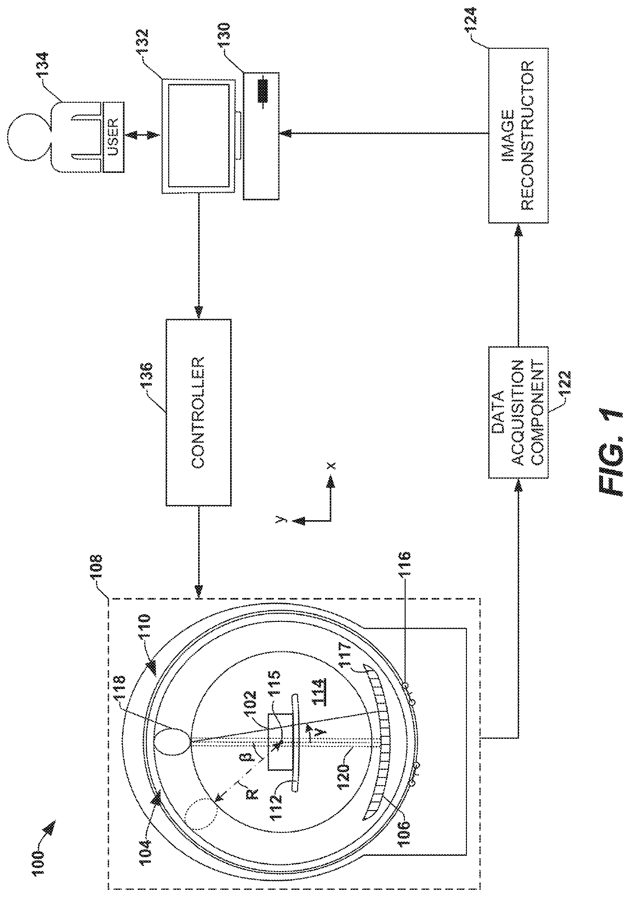
[0018]The claimed subject matter is now described with reference to the drawings, wherein like reference numerals are generally used to refer to like elements throughout. In the following description, for purposes of explanation, numerous specific details are set forth in order to provide a thorough understanding of the claimed subject matter. It may be evident, however, that the claimed subject matter may be practiced without these specific details. In other instances, structures and devices are illustrated in block diagram form in order to facilitate describing the claimed subject matter.

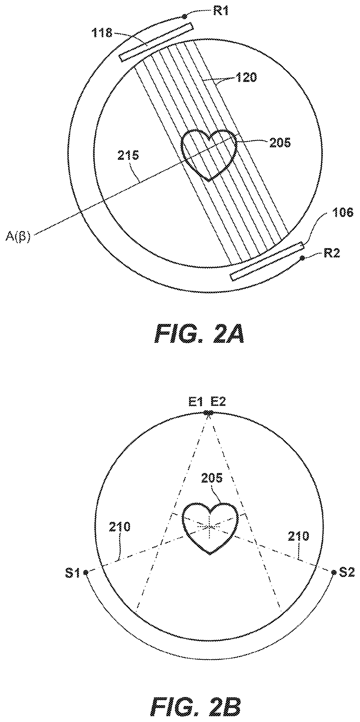
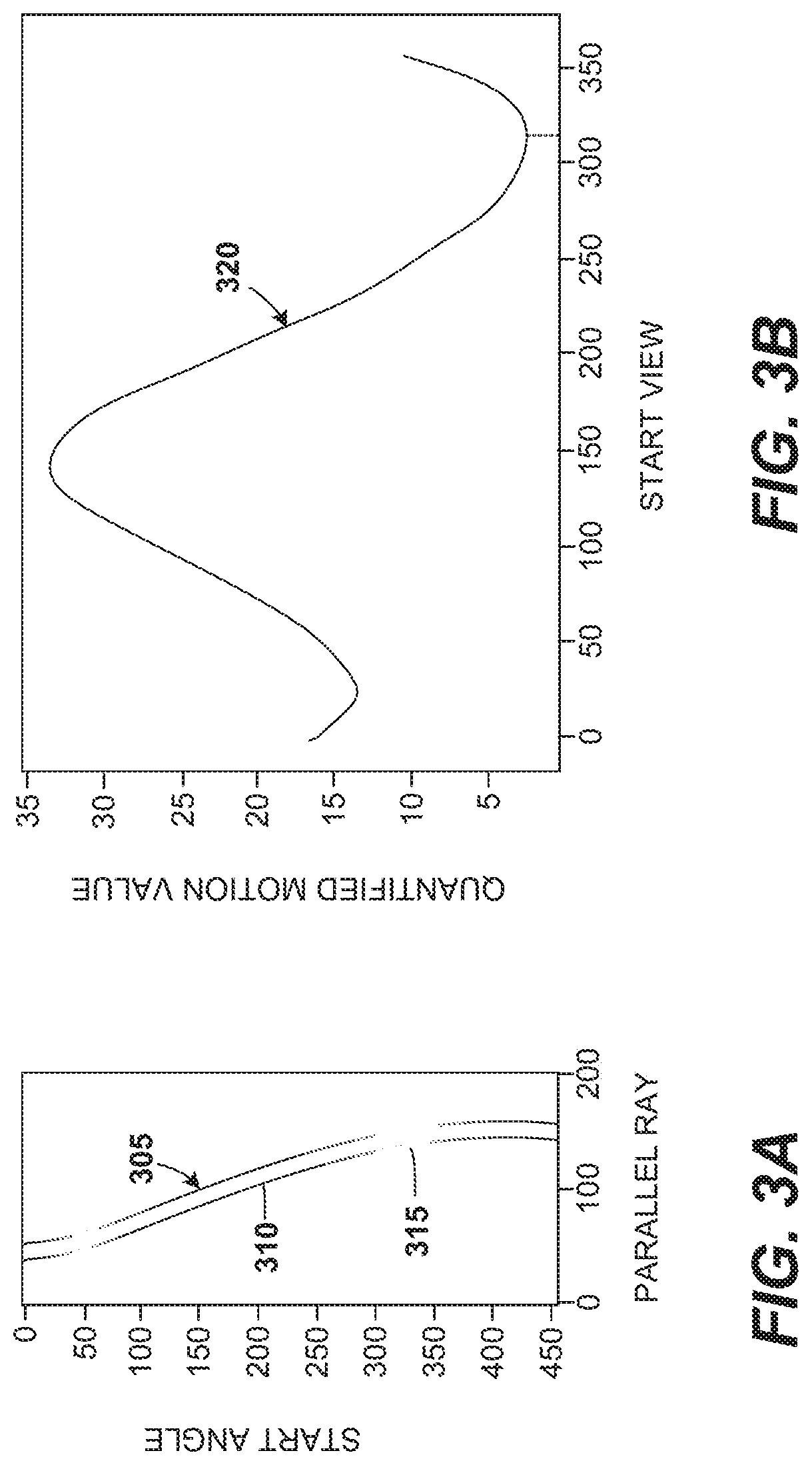
[0019]Among other things, one or more systems and / or techniques for mitigating motion artifacts in a computed tomography image of an anatomical object are provided herein. Anatomical objects such as a beating heart, for example, necessarily move when functioning properly. Movements of the heart during a heartbeat may seem to occur according to a fixed periodical schedule, but the timing of such mo...


PUM


Abstract

Provided are one or more systems and / or techniques for mitigating motion artifacts in a computed tomography image of an anatomical object. Extended scan data is received and includes projections and backprojections acquired for parallel rays emitted by a radiation source at different angular locations within a first range of source angles. The projections and the backprojections are compared to identify differences between the projections and the backprojections at the different angular locations. Movement of the anatomical object during acquisition of the extended scan data at the different angular locations is quantified, and short scan data is identified. The short set includes a subset of the extended scan data acquired at different locations within a second range of source angles where the quantified movement of the anatomical object is less than a movement threshold. The computed tomography image of the anatomical object is reconstructed from the short scan data.

Description

CROSS-REFERENCE TO RELATED APPLICATIONS[0001]This application is a national phase entry under 35 U.S.C. § 371 of International Patent Application PCT / US2018 / 0141777, filed Jan. 18, 2018, designating the United States of America, which claims the benefit under Article 8 of the Patent Cooperation Treaty to U.S. Provisional Patent Application Ser. No. 62 / 618,216, filed Jan. 17, 2018, for “Optimal Cardiac Phase in Prospectively Gated Axial Cardiac CT Scans,” the disclosure of each of which is hereby incorporated herein in its entirety by this reference.TECHNICAL FIELD[0002]The present application relates to the field of computed tomography (CT) and, more specifically, to image reconstruction systems and methods that at least partially eliminate motion artifacts from axially-aligned CT scans of a beating heart.BACKGROUND[0003]To improve temporal resolution in axial cardiac scans, short scans (e.g., half-scans, partial scans, etc.) are often used to obtain enough image data to allow a ful...

Claims


Application Information

Patent Timeline
IPC IPC(8): A61B6/03A61B6/00G06T11/00
CPCA61B6/5264A61B6/5211A61B6/486G06T2211/412G06T7/0012A61B6/5288G06T11/008A61B6/563G06T11/005G06T2207/10081A61B6/032A61B6/4064A61B6/503A61B6/5205A61B6/5258G01N23/046G01N2223/419G06T2207/10072G06T2211/404Y10S378/901
Inventor ZAMYATIN, ALEXANDERKARBEYAZ, BASAK ULKER
Owner ANLOGIC CORP (US)
Learn More