
Biological Tissue Access and Closure Apparatus, Systems and Methods

a biological tissue and apparatus technology, applied in the field of apparatus, systems and methods for accessing internal structures and closing biological tissue, can solve the problems of requiring considerable time to execute, difficult to perform most, if not all, current trocar access portal closing methods, etc., and achieve the effect of facilitating entry of such

Active Publication Date: 2019-12-05
SUTURE EASE
View PDF0 Cites 3 Cited by
  • Summary
  • Abstract
  • Description
  • Claims
  • Application Information

AI Technical Summary

Benefits of technology

The patent is about a device called a cannula lumen that can hold a suture guide and allows surgical instruments to enter the body during a surgery. The cannula lumen has a specific size and shape that is designed to fit the suture guide. This device helps make surgeries safer and easier.

Problems solved by technology

It is believed that inadequate closure of the trocar access portal is the primary cause of trocar-site hernias.
Although the noted methods can, in most instances, be employed to successfully close a trocar access portal, there are several significant drawbacks and disadvantages associated with the methods.
Indeed, as discussed below, most, if not all, current trocar access portal closing methods are typically difficult to perform, require considerable time to execute and do not provide for a simple, reproducible and reliable means of closing the trocar access portal.
A major drawback and disadvantage associated with the disclosed methods is that the apparatus and methods are primarily dependent on the dexterity of the surgeon operating the apparatus and executing the associated methods.
A further disadvantage associated with the methods disclosed in U.S. Pat. Nos. 919,138, 3,946,740 and 4,621,640 is that the surgeon operating the apparatus associated with the methods must either perform the trocar access portal closure “blind”, i.e. without visual access to interior body tissues, e.g., intra-abdominal fascia, or with the assistance of an endoscope inserted into the body cavity from an additional access portal.
A further disadvantage is that an exposed needle must be handled by a surgeon inside of a body cavity with limited visual access, thus, which increases the risk of injury to the local structures inside of a patient, e.g., organs.
A further disadvantage associated with the apparatus and methods disclosed in U.S. Pat. Nos. 919,138, 3,946,740 and 4,621,640 is that, if the surgeon desires to place more than one suture throw through the tissue, the surgeon must reload the needle into a needle driver apparatus.
This process is time consuming and oftentimes a frustrating exercise in hand-to-eye coordination.
Another drawback associated with the apparatus and associated methods disclosed in U.S. Pat. Nos. 919,138, 3,946,740 and 4,621,640 is that the apparatus can, and often times will, fail to effectively close tissue that is disposed proximate the trocar access portal, which greatly increases the patient's risk of trocar-site herniation at the closure site.
The seminal complications associated with trocar-site herniation include organ necrosis and closed loop intestinal obstruction, which can be life-threatening.
A further drawback is that patients with relatively thick body tissues increase the difficulty, time and risk of trocar access portal closure complications, such as a misplaced suture and / or penetration of a patient's organs with the suture needle.
A major drawback and disadvantage associated with the methods disclosed in U.S. Pat. No. 8,109,943 and Pub.
No. 2016 / 0228107 is that the stitch produced does not encompass the anterior fascia and peritoneum and, hence, does not fully close the defect in a traditional manner.
A further drawback associated with the methods disclosed in U.S. Pat. No. 8,109,943 and Pub.
No. 2016 / 0228107 is that, even if the trocar device is appropriately positioned in a patient, there is no means to ensure that the anchors will completely penetrate the targeted body tissues and successfully close the trocar access portal.
A further drawback is that at least two (2) exposed suture needles must be handled by a surgeon inside of a patient's body cavity with limited visual aid, which, as indicated above, greatly increases the risk of injury to the local structures, e.g., organs.
Another drawback associated with the apparatus and methods disclosed in U.S. Pat. No. 8,109,943 and Pub.
The suture anchors thus can, and often times will, elicit an adverse inflammatory response in the patient.
There is also a substantial risk of dislodgment of the suture anchors from body tissue, which can also cause serious postoperative complications.
Several drawbacks and disadvantages are similarly associated with the Medtronic® device and associated method.
A major drawback and disadvantage is that there is no reliable means associated with the Medtronic® device for a surgeon to assess whether the device is properly positioned in (or through) the body tissue of a patient.
It is, however, very difficult, if not impossible to achieve proper trocar device positioning for every patient via the fixed circumferential bands due to varying body tissue thicknesses encountered from patient-to-patient.
As a result of inaccurate positioning of the Medtronic® device, the associated method can cause unintended damage or trauma to local tissues.
The Medtronic® method can also fail to successfully close a trocar access portal, which can lead to the above noted complications, e.g., trocar-site herniation.
A further drawback associated with the Medtronic® method is that the method requires a secondary grasper tool controlled from an additional trocar access portal in order to guide a suture through the two (2) contra-laterally opposed regions of body tissue.
A further drawback associated with the Medtronic® method is that it is extremely difficult and cumbersome to manipulate and engage the intra-cavity suture with the grasper tool.
Another drawback associated with the Medtronic® method is that the method also requires that at least one (1) sharp instrument be introduced to and manipulated within a body cavity with limited visual access to interior body tissues, which as indicated above, substantially increases the risk of injury to the local structures inside of a patient, e.g., organs.

Method used


Image

  • Biological Tissue Access and Closure Apparatus, Systems and Methods
  • Biological Tissue Access and Closure Apparatus, Systems and Methods
  • Biological Tissue Access and Closure Apparatus, Systems and Methods

Examples


Embodiment Construction

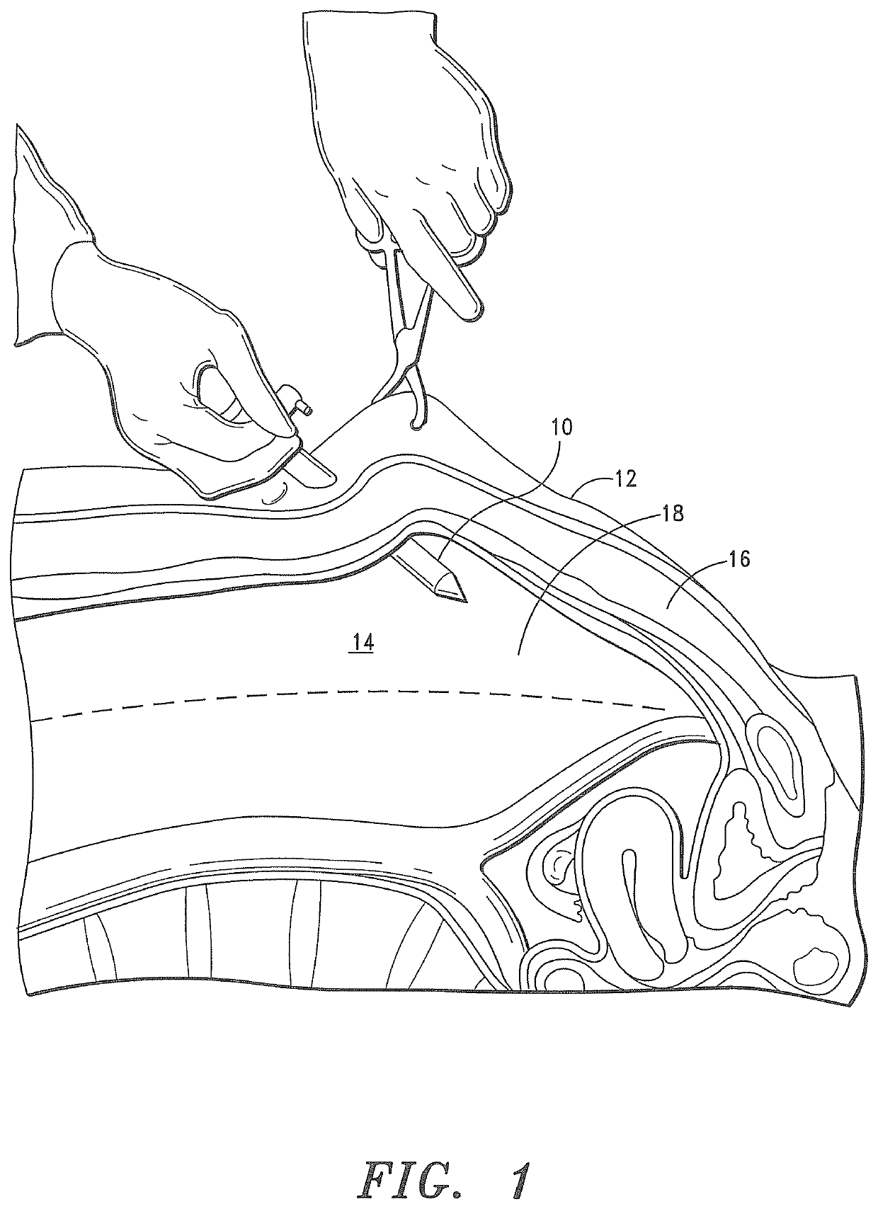
[0087]Before describing the present invention in detail, it is to be understood that this invention is not limited to particularly exemplified apparatus, systems, structures or methods as such may, of course, vary. Thus, although a number of apparatus, systems and methods similar or equivalent to those described herein can be used in the practice of the present invention, the preferred apparatus, systems, structures and methods are described herein.

[0088]It is also to be understood that, although the present invention is described and illustrated in connection with laparoscopic procedures, the invention is not limited to such procedures. According to the invention, the apparatus, systems and methods of the invention can also be employed in connection with a multitude of other surgical procedures, including, without limitation, patent foramen ovale (PFO) closure, left ventricular closure and vascular, i.e. artery and vein, closure.

[0089]It is also to be understood that the terminolog...


PUM


Abstract

A system for piercing through biological tissue and providing access to internal structures; particularly, intra-abdominal structures, and closing an opening in the tissue. The system includes a suture guide sub-system having a tissue positioning sub-system for engaging segments of tissue disposed proximate the tissue opening and a suture passer sub-system that slideably engages the suture guide sub-system. The suture passer sub-system includes suture deployment and capture means that is configured to removably secure a suture thereto and deliver the suture to the segments of tissue disposed proximate the tissue opening.

Description

CROSS-REFERENCES TO RELATED APPLICATIONS[0001]This application is a continuation-in-part of U.S. application Ser. No. 15 / 991,441, filed on May 29, 2018, which claims the benefit of U.S. Provisional Application No. 62 / 512,180, filed on May 30, 2017, U.S. Provisional Application No. 62 / 618,634, filed on Jan. 18, 2018, and U.S. Provisional Application No. 62 / 655,151, filed on Apr. 9, 2018.FIELD OF THE INVENTION[0002]The present invention relates to apparatus, systems and methods for accessing internal structures and closing biological tissue. More particularly, the present invention relates to apparatus, systems and methods for accessing internal structures; particularly, intra-abdominal structures, and approximation, ligation, fixation and closure of openings in biological tissue; particularly, laparoscopic ports or incisions in biological tissue.BACKGROUND OF THE INVENTION[0003]As reported in a Jan. 31, 2017 iData Research press release, approximately 3.5 million laparoscopic surgica...

Claims


Application Information

Patent Timeline
Patent Type & Authority Applications(United States)
IPC IPC(8): A61B17/04A61B17/34
CPCA61B17/0491A61B17/0482A61B17/3423A61B17/0469A61B2017/3447A61B2017/00637A61B2017/00663A61B2017/06042A61B2017/06009A61B2017/0472A61B17/3421A61B17/3417
Inventor HENEVELD, SCOTT
Owner SUTURE EASE
Learn More