
Catadioptric medical imaging system for observing the inside wall of a surgical cavity

Active Publication Date: 2019-09-12
LEICA INSTR SINGAPORE PTE
View PDF0 Cites 0 Cited by
  • Summary
  • Abstract
  • Description
  • Claims
  • Application Information

AI Technical Summary

Benefits of technology

The patent describes a system that uses a convex catoptric mirror to provide a surgeon with a clear view of the interior of a surgical cavity during a surgical procedure. A camera device records the image of the interior wall of the cavity through the mirror. The patent also includes a correction module with optics that can correct distortion in the mirror image, allowing the surgeon to have a clear and accurate view of the cavity.

Problems solved by technology

During surgery, it is often difficult for the surgeon to gain information about the structure of a surgical cavity, in particular if the surgical cavity widens and forms an undercut, which cannot be observed in a front view.

Method used


Image

  • Catadioptric medical imaging system for observing the inside wall of a surgical cavity

Examples


Embodiment Construction

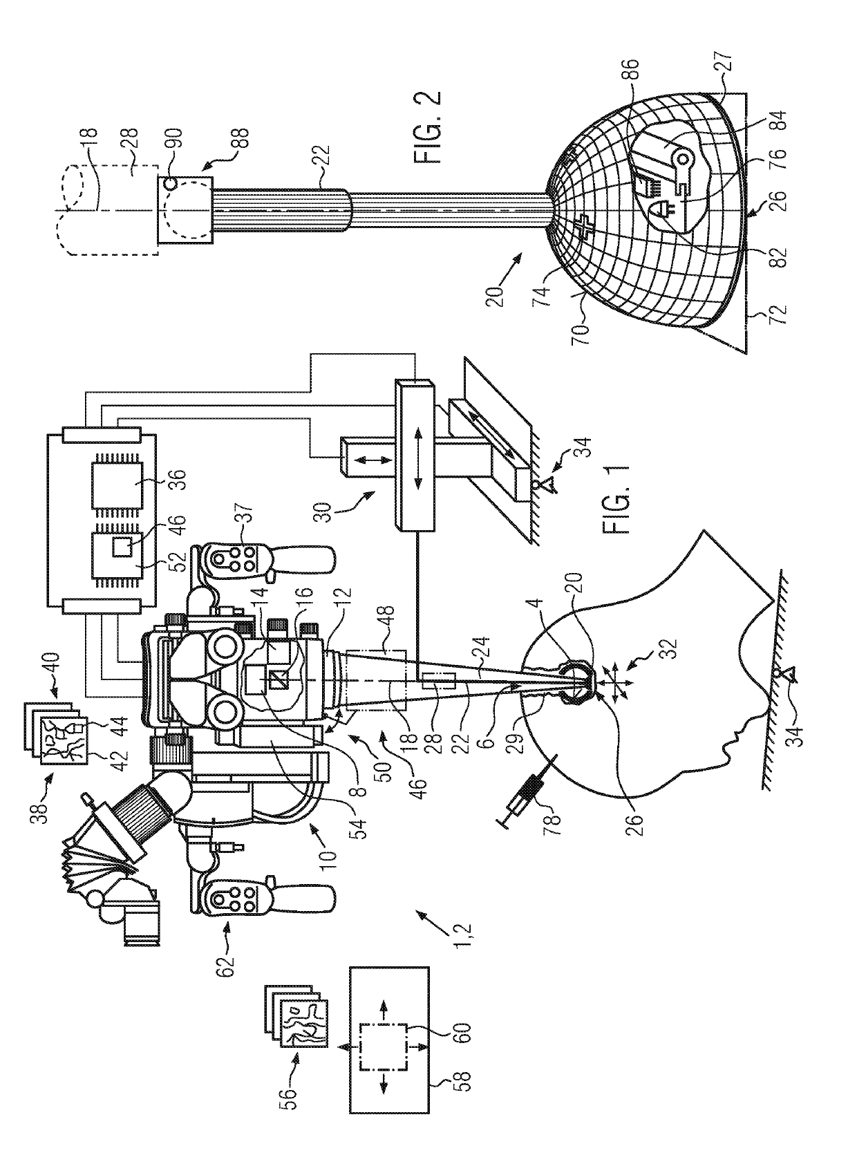
[0046]First, the design and function of a catadioptric medical imaging system 1, such as a surgical microscope 2 are explained with reference to FIG. 1. The catadioptric medical imaging system 1 is used for observing an inside wall 4 of a surgical cavity 6, such as a surgical cavity in brain surgery, but also for any other kind of surgical cavity.

[0047]The catadioptric medical imaging system 1 comprises a camera device 8 which may be located in an optics carrier 10 together with a lens 12, in particular a microscope lens. The catadioptric medical imaging system 1 may also comprise an illumination device 14 which may also be included in the optics carrier 10. A beam splitter 16 may be provided to add light from the illumination device 14 coaxial to an optical axis 18 of the camera device 8 and directed towards the surgical cavity 6. Light directed towards the camera device 8 from the surgical cavity 6 is separated from light from the illumination device 14 by the beam splitter 16.

[00...


PUM


Abstract

The invention relates to a catadioptric medical imaging system (1), in particular a surgical microscope (2). During surgery, it may be necessary to gain more information about a surgical cavity (6), in particular the type of tissue (29) at the inside walls (4) of the surgical cavity (6). To solve this problem, the catadioptric medical imaging system (1) according to the invention comprises a camera device (8) and a convex catoptric mirror (20) adapted to be inserted into the surgical cavity (6). The catoptric mirror (20) is mounted on an arm (22) and spaced apart from the camera device (8).

Description

CROSS REFERENCE TO RELATED APPLICATIONS[0001]This application claims priority of European patent application number 18160210.3 filed Mar. 6, 2018, the entire disclosure of which is incorporated by reference herein.FIELD OF THE INVENTION[0002]The invention relates to a catadioptric medical imaging system for observing the inside wall of a surgical cavity.BACKGROUND OF THE INVENTION[0003]During surgery, it is often difficult for the surgeon to gain information about the structure of a surgical cavity, in particular if the surgical cavity widens and forms an undercut, which cannot be observed in a front view.SUMMARY OF THE INVENTION[0004]The object of the present invention is to provide a system and a method for obtaining such information.[0005]This object is achieved according to the invention in that the catadioptric medical imaging system for observing the inside wall of a surgical cavity comprises a camera device and a convex catoptric mirror adapted to be inserted into the surgica...

Claims


Application Information

Patent Timeline
IPC IPC(8): G02B17/08A61B90/00G02B21/00
CPCG02B17/0856G02B17/023G02B21/0012A61B5/0075A61B90/361G02B17/0804G02B17/0892G02B21/02A61B90/30A61B2090/309A61B2090/3618A61B90/20A61B2090/371A61B2090/372A61B2090/3941G02B13/06G02B17/08G02B21/025G02B21/04
Inventor THEMELIS, GEORGE
Owner LEICA INSTR SINGAPORE PTE
Learn More