Eureka AIR delivers breakthrough ideas for toughest innovation challenges, trusted by R&D personnel around the world.

Method for producing anatomical models and models obtained

a technology of anatomical models and models, applied in the field of medicine, can solve the problems of limiting the production of soft organ models, -hardness limitations of rigid 3d printing materials, and -inability to produce anatomical models of soft organs such as livers or breasts, and achieve the effect of ensuring the consistency of the actual organ

Inactive Publication Date: 2018-12-06
CELLA MEDICAL SOLUTIONS SL
View PDF0 Cites 9 Cited by
  • Summary
  • Abstract
  • Description
  • Claims
  • Application Information

AI Technical Summary

Benefits of technology

The invention is about a new method for making anatomical models of soft organs, such as the liver or breast, and even specific patients' models. The method involves scanning the organ using image diagnostic techniques and using software to select and segment the anatomical structure of interest. The model is then made using 3D printing technology, using rigid or semi-rigid materials to replicate the internal parts of the organ and soft materials like silicones or silicone gels to represent the outer part. The model can be used for educational and training purposes and allows for emulating surgical procedures. The method is cost-effective and allows for customization for each patient.

Problems solved by technology

However, direct printing of the part limits the production of soft organ models, since the non-rigid 3D printing materials have hardness limitations and are expensive.
In this way, anatomical models of soft organs, such as liver or breast, would not be possible with the actual consistency of the organ.
It is only possible to obtain hard materials without the possibility of emulating surgical cutting and suturing procedures, or with minimum hardness levels of “shore A27”, which is not sufficient to emulate the consistency and deformation of these organs.

Method used


Image

  • Method for producing anatomical models and models obtained
  • Method for producing anatomical models and models obtained

Examples


Embodiment Construction

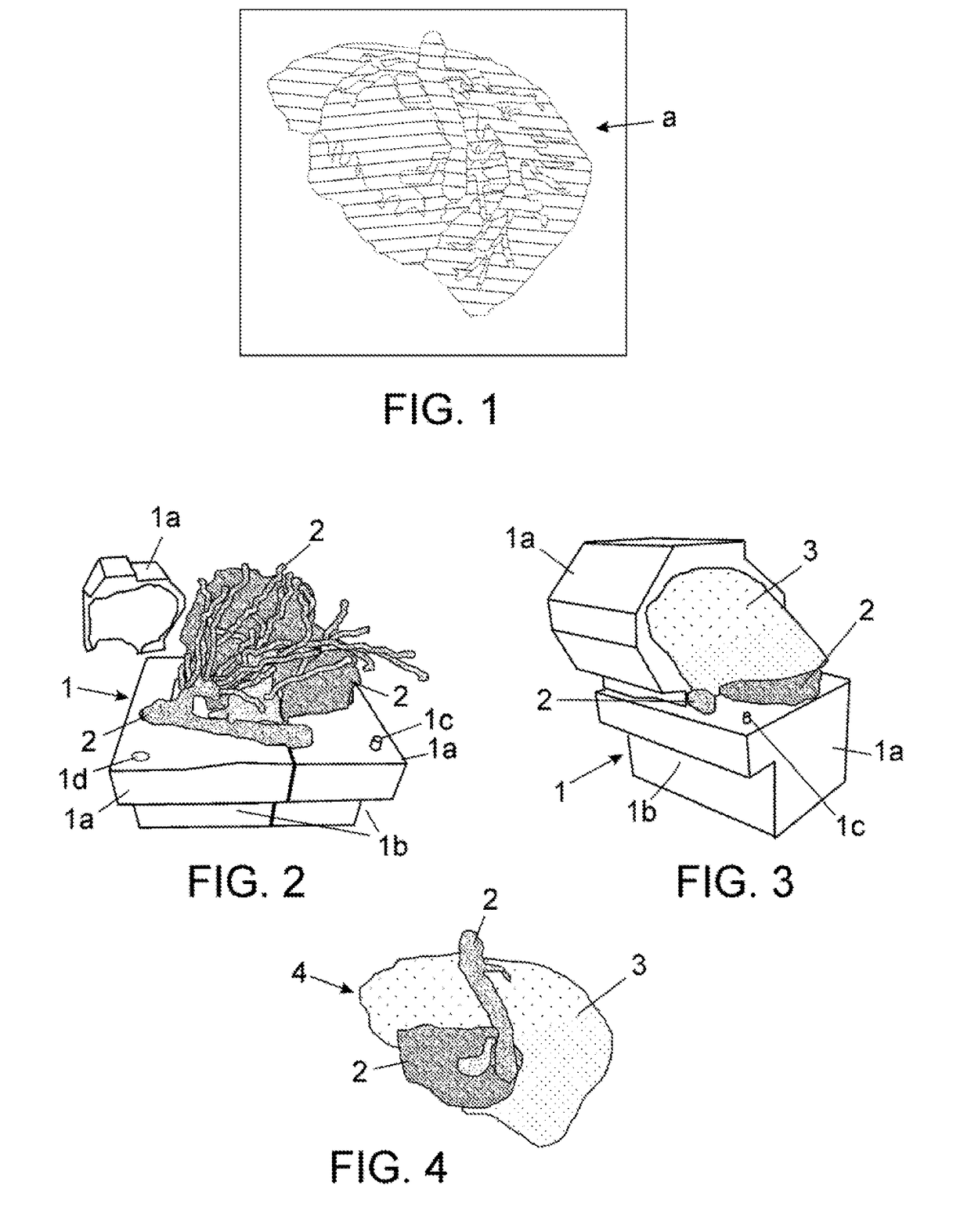
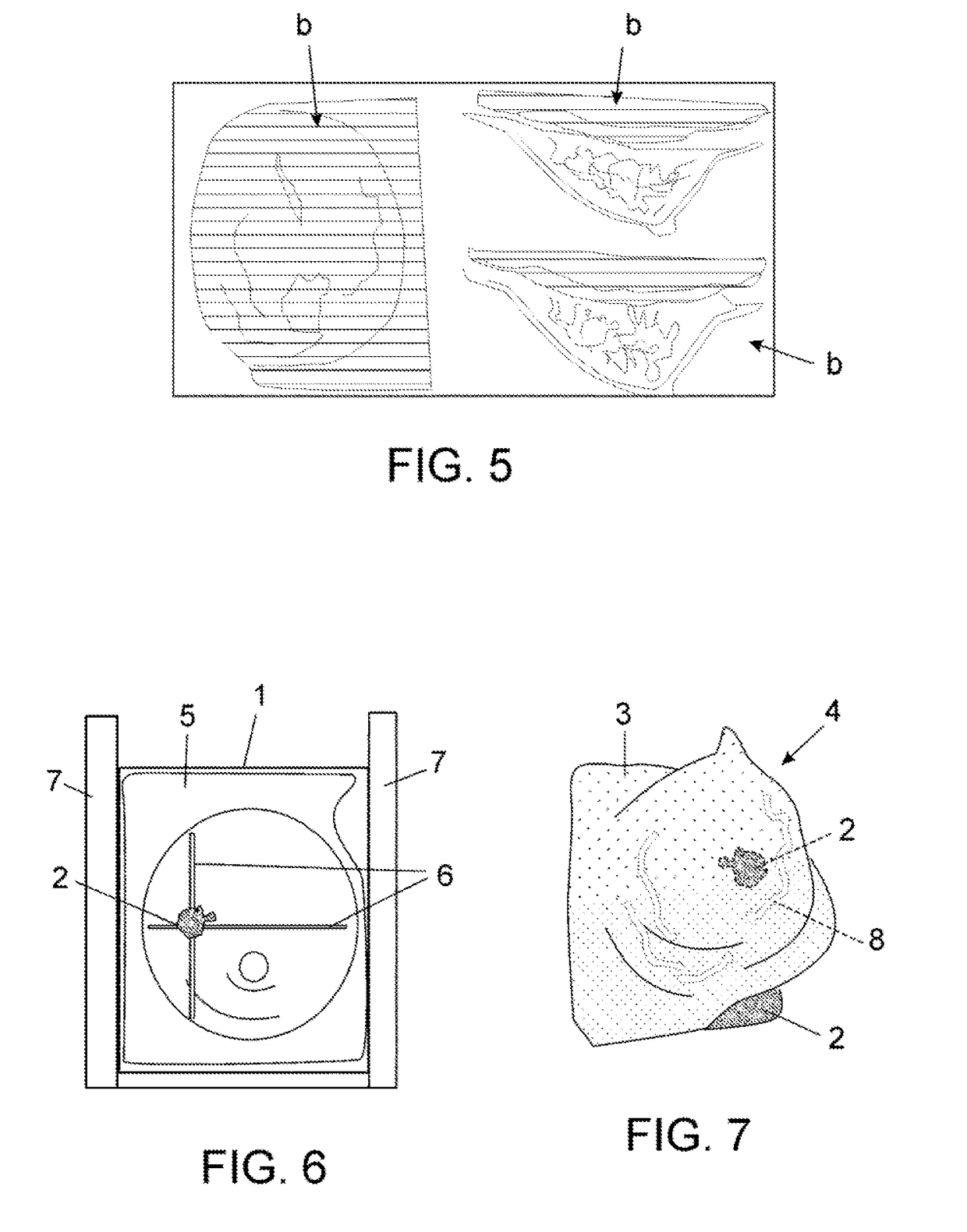
[0031]In view of the aforementioned figures, and according to their numbering, a non-limiting exemplary embodiment of the method for the manufacture of anatomical liver models (A) and breast (B) can be seen, which comprises the following:

Example (A) for Anatomical Models of Liver

[0032]The method of making anatomical models of liver comprises, in the first stage, obtaining information about the patient's liver by an image diagnosis such as a CAT (Computerized Axial Tomography) with or without vascular reconstruction, NMR (Nuclear Magnetic Resonance), Echography, Cholangiography or the like.

[0033]Next, in a second stage, specialized software has been developed for selecting (segmenting) automatically different elements of the organ; in particular, the following elements in the images obtained: the liver parenchyma, hepatic-biliary vasculature differentiating each of the elements and tumor (when appropriate). It will be apparent that other programs known to those skilled in the art may...


PUM


Abstract

A method for producing anatomical models and models obtained are disclosed. The method includes obtaining information by diagnostic imaging; obtaining a three-dimensional, computerised model of the anatomical structure; and performing the following steps: designing a negative mould; printing the negative mould in 3D; printing in 3D rigid pieces of internal elements of the model provided; placing the pieces in the mould; closing and sealing the mould; injecting a soft material into the mould; and removing same from the mould. The anatomical model is a liver with hepatobiliary vasculature and tumours made from the rigid parts and hepatic parenchyma made from the soft material, or it is a mammary gland with a tumour and muscular tissue made from the rigid parts, mammary tissue made from the soft material and an external covering as skin.

Description

OBJECT OF THE INVENTION[0001]The invention, as expressed in the title of the present specification, relates to a method for manufacturing anatomical models and to the obtained models, providing features, which will be described in detail below, which involve a novelty in the current state of the art within its field of application.[0002]More particularly, the object of the invention is directed to a method of making anatomical models of human organs, or parts thereof. In particular, soft organs, such as liver or breast models, which have utility in teaching and different disciplines of the medical sector, such as the planning and simulation of surgeries, incorporating significant innovations and advantages over current manufacturing processes thereof. A second aspect of the invention is the same anatomical models obtained from such manufacturing process.FIELD OF APPLICATION OF THE INVENTION[0003]The application field of this invention is marked within the field of medicine. The inve...

Claims


Application Information

Patent Timeline
IPC IPC(8): G09B23/30A61F2/30A61B34/10
CPCG09B23/30A61F2/30942A61B34/10A61B2034/102G05B19/4099A61F2002/30952G06T2207/10081G06T2207/30056B33Y80/00G06T2210/41B33Y50/02A61B2034/105G09B23/34
Inventor GARC A CALDERON, DARIO
Owner CELLA MEDICAL SOLUTIONS SL
Why Eureka
Eureka Blog
Learn More