
Digital histopathology and microdissection

a microdissection and histopathology technology, applied in the field of histopathology and microscopic examination of tissue, can solve the problems of unreliability, time-consuming, expensive,

Active Publication Date: 2018-04-26
NANTOMICS LLC
View PDF1 Cites 39 Cited by
  • Summary
  • Abstract
  • Description
  • Claims
  • Application Information

AI Technical Summary

Benefits of technology

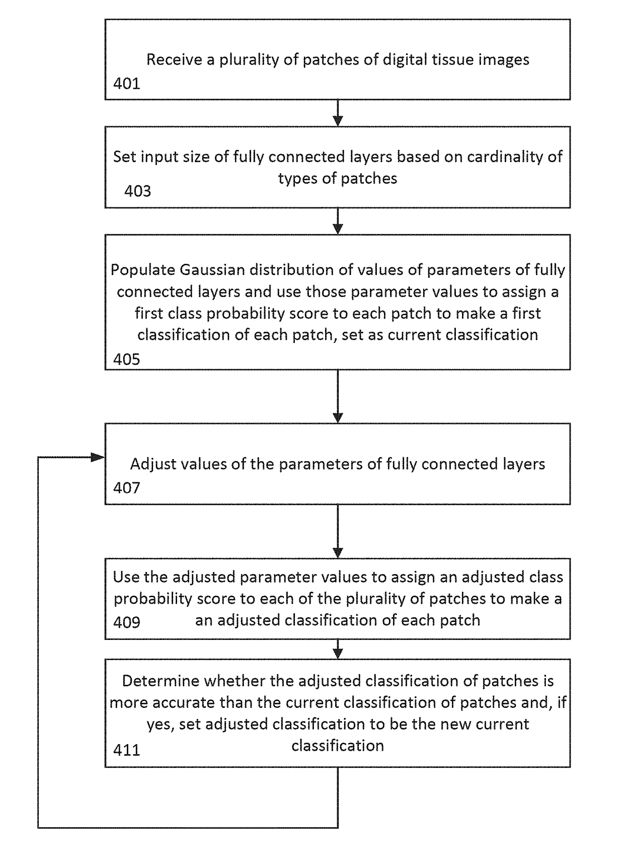
The present invention is about using neural networks to identify regions of interest in digital images of tissue. This technology can be used to determine whether tissue is likely to be diseased, which is currently a time-consuming and unreliable process. The invention aims to provide a solution to this problem by using convolutional neural networks to identify these regions of interest. The invention can be used in various settings, including biopsy, surgery, or autopsy, and can be applied to different types of tissue. The invention can also be used in combination with other techniques, such as verification by multiple pathologists to minimize false determinations. Overall, the invention provides a faster and more accurate way to determine if tissue is likely to be diseased.

Problems solved by technology

However, there is a technical problem with these determinations in that they are often unreliable, expensive, time consuming, and generally require verification by multiple pathologists to minimize the likelihood of false determinations, including false positives as well as false negatives.

Method used


Image

  • Digital histopathology and microdissection
  • Digital histopathology and microdissection
  • Digital histopathology and microdissection

Examples


Embodiment Construction

[0020]The present invention will now be described more fully hereinafter with reference to the accompanying drawings which show, by way of illustration, specific embodiments by which the invention may be practiced. This invention may, however, be embodied in many different forms and should not be construed as limited to the embodiments set forth herein; rather, these embodiments are provided so that this disclosure will be thorough and complete, and will fully convey the scope of the invention to those skilled in the art. Among other things, the present invention may be embodied as devices or methods. Accordingly, the present invention may take the form of an entirely hardware embodiment, an entirely software embodiment, or an embodiment combining software and hardware aspects. The following detailed description is, therefore, not to be taken in a limiting sense.

[0021]Throughout the specification and claims, the following terms take the meanings explicitly associated herein, unless ...


PUM


Abstract

A computer implemented method of generating at least one shape of a region of interest in a digital image is provided. The method includes obtaining, by an image processing engine, access to a digital tissue image of a biological sample; tiling, by the image processing engine, the digital tissue image into a collection of image patches; identifying, by the image processing engine, a set of target tissue patches from the collection of image patches as a function of pixel content within the collection of image patches; assigning, by the image processing engine, each target tissue patch of the set of target tissue patches an initial class probability score indicating a probability that the target tissue patch falls within a class of interest, the initial class probability score generated by a trained classifier executed on each target tissue patch; generating, by the image processing engine, a first set of tissue region seed patches by identifying target tissue patches having initial class probability scores that satisfy a first seed region criteria, the first set of tissue region seed patches comprising a subset of the set of target tissue patches; generating, by the image processing engine, a second set of tissue region seed patches by identifying target tissue patches having initial class probability scores that satisfy a second seed region criteria, the second set of tissue region seed patches comprising a subset of the set of target tissue patches; calculating, by the image processing engine, a region of interest score for each patch in the second set of tissue region seed patches as a function of initial class probability scores of neighboring patches of the second set of tissue region seed patches and a distance to patches within the first set of issue region seed patches; and generating, by the image processing engine, one or more region of interest shapes by grouping neighboring patches based on their region of interest scores.

Description

CROSS REFERENCE TO RELATED APPLICATIONS[0001]This application claims the benefit of priority to U.S. Provisional Application No. 62 / 411,290, filed Oct. 21, 2016, and U.S. Provisional Application No. 62 / 557,737, filed Sep. 12, 2017. These and all other extrinsic materials referenced herein are incorporated by reference in their entirety.INTRODUCTION[0002]The present technology relates generally to histopathology, the microscopic examination of tissue for the purpose of determining whether the tissue is diseased and / or studying diseased tissue. The tissue may be removed from any part of the body including, for example, breast lumps, specimens of bowel, kidney, liver, uterus lining, lung, chest, lymph node, muscle, nerve, skin, testicle, thyroid, or the like.[0003]This disclosed technology relates to identifying regions of interest within a digital image, for example, identifying foreground objects from background scenes, or identifying cancer cells within a digital histopathology imag...

Claims


Application Information

Patent Timeline
IPC IPC(8): G06T7/00G06K9/62G06N3/04G06V10/50G06V10/764
CPCG06T7/0012G06K9/6227G06K9/6223G06K9/6278G06K9/6276G06K9/6296G06K9/6269G06K9/6217G06N3/04G06T2207/30024G06N20/00G06T2207/10056G06T2207/20021G06T2207/20084G06T2207/20156G06T7/11G06T7/187G06V20/695G06V10/50G06V10/454G06V10/82G06V10/7796G06V10/764G06N7/01G06N3/045G06T1/20G06F18/21G06F18/29G06F18/285G06F18/2193G06F18/2411G06F18/23213G06F18/24137G06F18/24147G06F18/24155
Inventor SONG, BINGCHU, GREGORY
Owner NANTOMICS LLC
Learn More