
Method for laser printing biological components, and device for implementing said method

a biological component and laser printing technology, applied in the direction of manufacturing tools, applying layer means, skeletal/connective tissue cells, etc., can solve the problems of difficult to deal with the complexity of tissues, limited growth of biological tissues, and unsatisfactory first procedures, etc., to achieve better resolution, less cell density, and better resolution

Active Publication Date: 2017-11-09
UNIV DE BORDEAUX +1
View PDF9 Cites 13 Cited by
  • Summary
  • Abstract
  • Description
  • Claims
  • Application Information

AI Technical Summary

Benefits of technology

The patent text describes different printing methods for bio-components using laser technology. These methods allow for the production of complex biological tissues with high cell density and resolution. The combination of bio-extrusion and laser technology allows for higher production rates and more complex biological tissues. The printing device includes a nozzle print head for printing bio-inks and a laser-type print head to print the extracellular matrix and cells simultaneously. The device also includes a chamber for storing the donor substrate and maintaining an atmosphere suitable for bio-inks.

Problems solved by technology

This first procedure is not fully satisfactory because the growth of the biological tissue can be limited in the depth of the macroporous matrix due to a lack of cell colonization.
Besides, this procedure makes it difficult to deal with the complexity of the tissues (which is characterized by a multiplicity of cell types organized in specific and generally anisotropic arrangements).
Bio-inks with high cell density can thus be printed with difficulty, with a high resolution since such printing technique induces, upon the passage of ink through the nozzle, high shear stresses liable to damage the cells.
Besides, with this type of ink, the risk of nozzles being blocked by the cells is important mainly because of the settling of cells inside the tanks.
Even though laser printing theoretically makes it possible to reach a high printing rate, insofar as it is possible to generate several tens of thousands of droplets per second, the production of biological tissues is not fast since these droplets represent a few tens of picoliters only.
Even though this second embodiment uses donor substrates containing a larger volume of bio-ink, it is not necessarily suitable for bio-inks.
As the concentration in elements to be printed is low near the free surface, the printed droplets have de facto low concentrations of cells, which is generally detrimental to the printed biological tissue.
Besides, according to this method, the number of cells and the concentration of deposited cells can hardly be controlled.

Method used


Image

  • Method for laser printing biological components, and device for implementing said method
  • Method for laser printing biological components, and device for implementing said method
  • Method for laser printing biological components, and device for implementing said method

Examples


Embodiment Construction

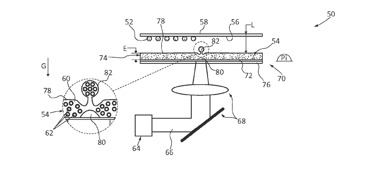
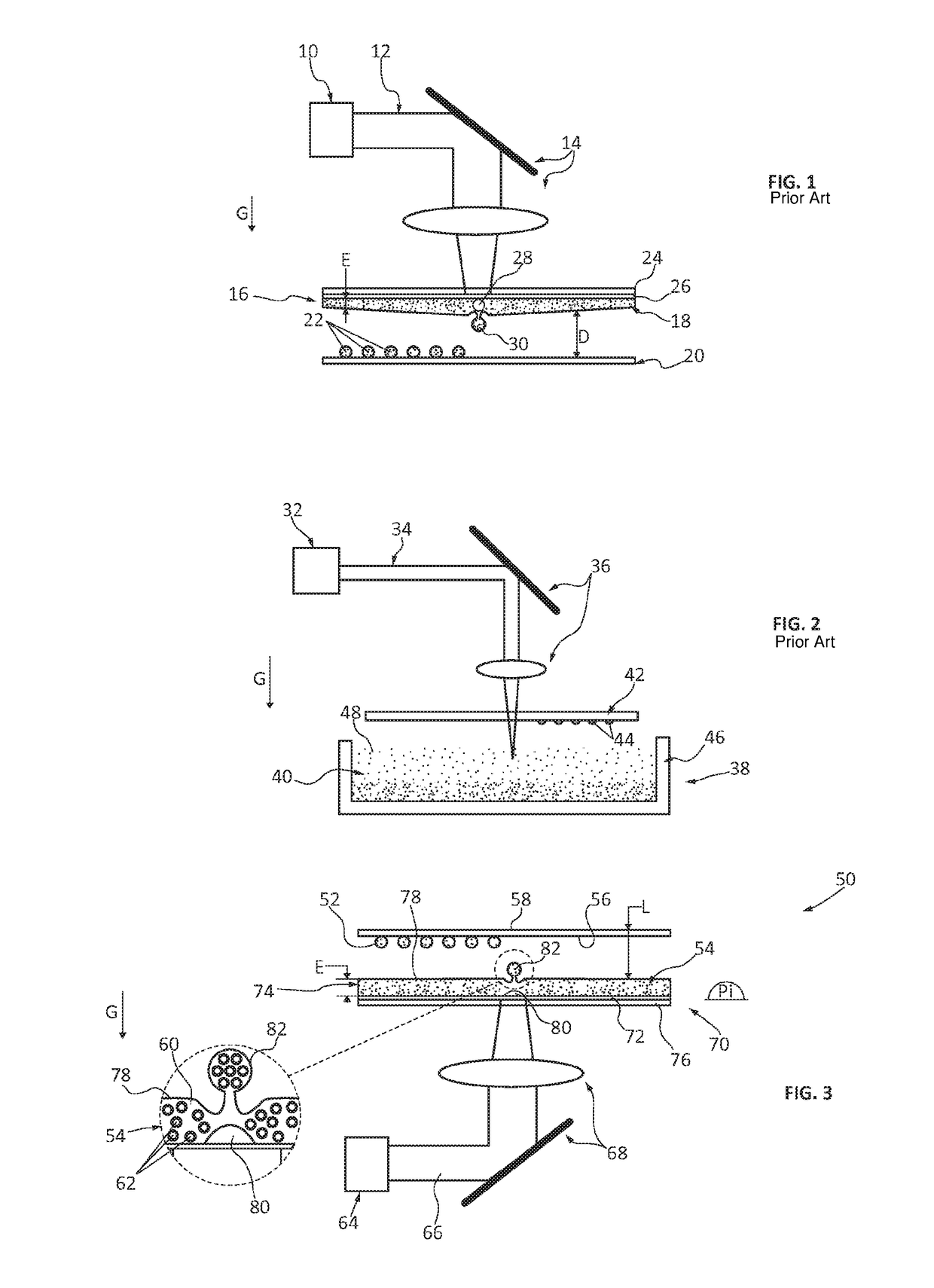
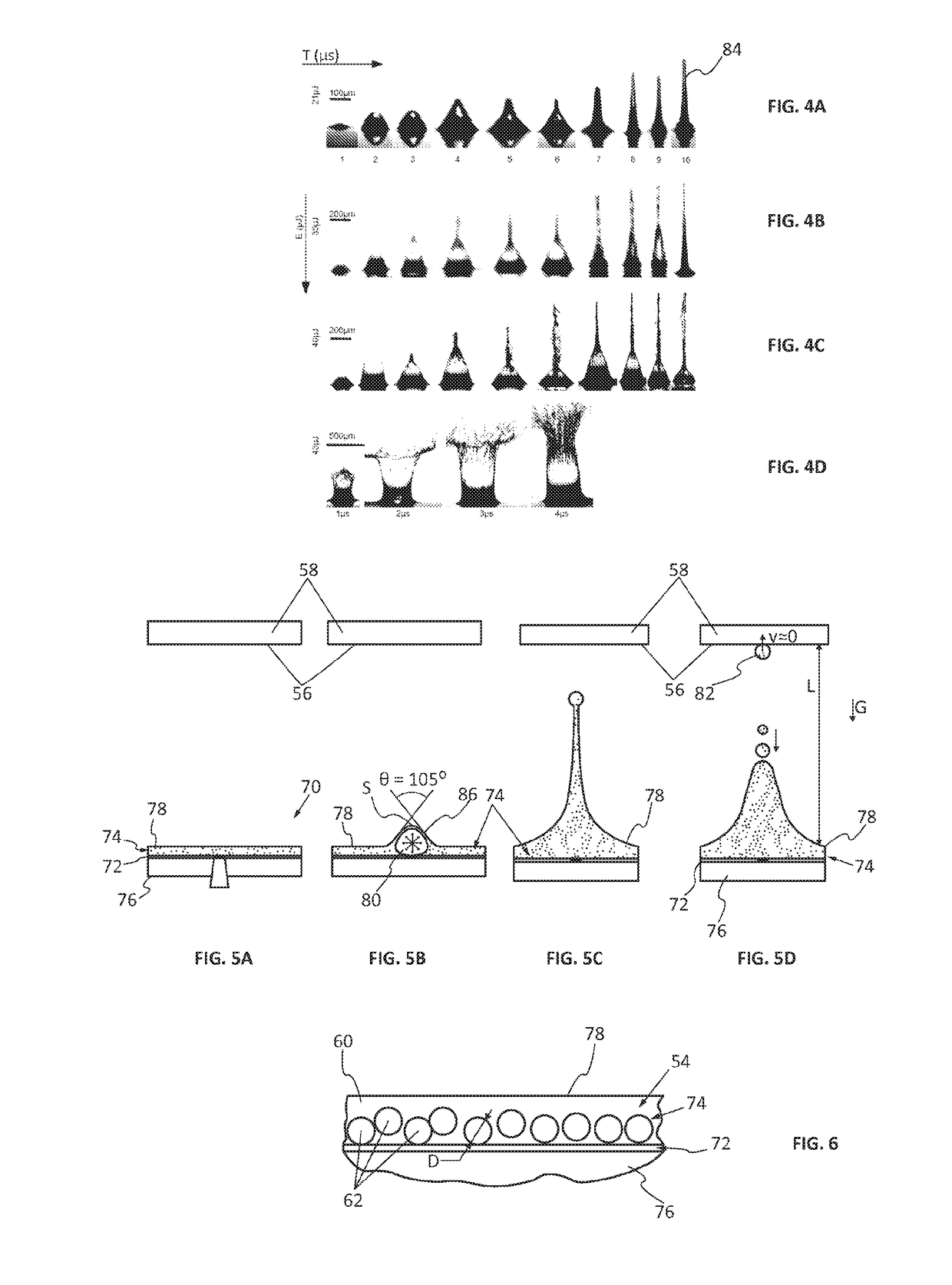
[0045]FIG. 3 shows a printing device 50 for producing at least one biological tissue through a layer by layer assembling according to a predefined arrangement, various components such as an extracellular matrix and various morphogens. Thus, the printing device 50 makes it possible to deposit, layer by layer, droplets 52 of at least one bio-ink 54 onto a depositing surface 56 which corresponds to the surface of a receiving substrate 58 for the first layer or for the last layer deposited on said receiving substrate 58 for the following layers. In order to simplify the representation, the depositing surface 56 corresponds to the surface of the receiving substrate 58 in FIG. 3.

[0046]According to one embodiment in FIG. 6, the bio-ink 54 comprises a matrix 60, for example an aqueous medium, wherein elements 62, for example, cells or cell aggregates, to be printed onto the depositing surface 56 can be found. As the case may be, a bio-ink 54 comprises, in the matrix 60, only one kind of ele...


PUM

PropertyMeasurementUnit
Thicknessaaaaaaaaaa
Coloraaaaaaaaaa
Sizeaaaaaaaaaa

Abstract

A method for printing uses at least one bio-ink. The method also uses at least one laser print head to deposit at least one droplet of at least one bio-ink onto a depositing surface of a receiving substrate. The printing method uses at least one nozzle print head to deposit at least one droplet of at least one bio-ink onto a depositing surface of the same receiving substrate as the laser print head.

Description

CROSS-REFERENCE TO RELATED APPLICATIONS[0001]This application is a National Phase Entry of International Patent Application Serial Number PCT / FR2015 / 053570, filed on Dec. 17, 2015, which claims priority to French Patent Application Serial No. 14 / 62570, filed on Dec. 17, 2014, both of which are incorporated by reference herein.BACKGROUND AND SUMMARY[0002]The present invention relates to a method for laser printing biological components and to a device for the implementation thereof. The invention also relates to a method for producing a biological tissue using this printing method.[0003]According to a first procedure, cells can be grown in vitro on a biodegradable macroporous matrix (or scaffold) for obtaining a biological tissue. This first procedure is not fully satisfactory because the growth of the biological tissue can be limited in the depth of the macroporous matrix due to a lack of cell colonization. Besides, this procedure makes it difficult to deal with the complexity of th...

Claims


Application Information

Patent Timeline
IPC IPC(8): B29C64/112C12M1/00C12M3/00B33Y30/00B33Y10/00B29C64/209B33Y80/00C12N5/077B29C64/273B33Y70/00
CPCB29C64/112B29C64/273B33Y10/00B33Y30/00B29C64/209B33Y70/00C12M23/50C12M21/08C12N5/0654B29K2995/0056B33Y80/00C12M33/00B41J2/14104C12N2533/90C12N5/0068
Inventor GUILLEMOT, FABIEN
Owner UNIV DE BORDEAUX
Learn More