
Methods for coating implant surfaces to treat surgical infections

a technology of surgical infection and implant surface, which is applied in the direction of prosthesis, internal osteosynthesis, osteosynthesis devices, etc., can solve the problems of significant probability of reinfection and inability to successfully treat device-related infections with systemic antibiotics alon

Inactive Publication Date: 2017-06-22
BIOMET MFG CORP
View PDF6 Cites 12 Cited by
  • Summary
  • Abstract
  • Description
  • Claims
  • Application Information

AI Technical Summary

Benefits of technology

The patent is about a new type of joint prosthesis that does not require cement to be used during the installation process. This prosthesis is easier to revise and can be used for both initial joint repairs and revision procedures. The technical effects are that it reduces the risk of complications and associated costs associated with a two-stage revision process.

Problems solved by technology

Such device-related infections may not be successfully treated by systemic antibiotics alone.
Because the “cure” rate associated with a one-stage procedure is typically between 60-80%, there is a significant probability that reinfection will occur, requiring yet another revision surgery.

Method used


Image

  • Methods for coating implant surfaces to treat surgical infections
  • Methods for coating implant surfaces to treat surgical infections

Examples


example 1

[0072]0.005 g of rifampin and 0.005 g of minocycline are dissolved in 0.05 g of ethanol to form an antimicrobial mixture. About half of the ethanol is allowed to evaporate to form a concentrated antimicrobial mixture, which is then stirred into 0.1 g of Phosal® 53 MCT (Lipoid Group, Köln, Germany) until a uniform antimicrobial and lecithin mixture is formed. The antimicrobial / lecithin mixture is then folded into 10 g of Phospholipon® 90G (Lipoid Group, Köln, Germany) until a uniform composition is formed. The final composition contains:

[0073]0.1% each of rifampin and minocycline;

[0074]0.5% ethanol;

[0075]1% of a mixture of 50% phosphatidylcholine and 50% various other lipids; and

[0076]98.4% of a mixture of at least 90% phosphatidyl choline and the remainder a mixture of various other smaller lipids.

example 2

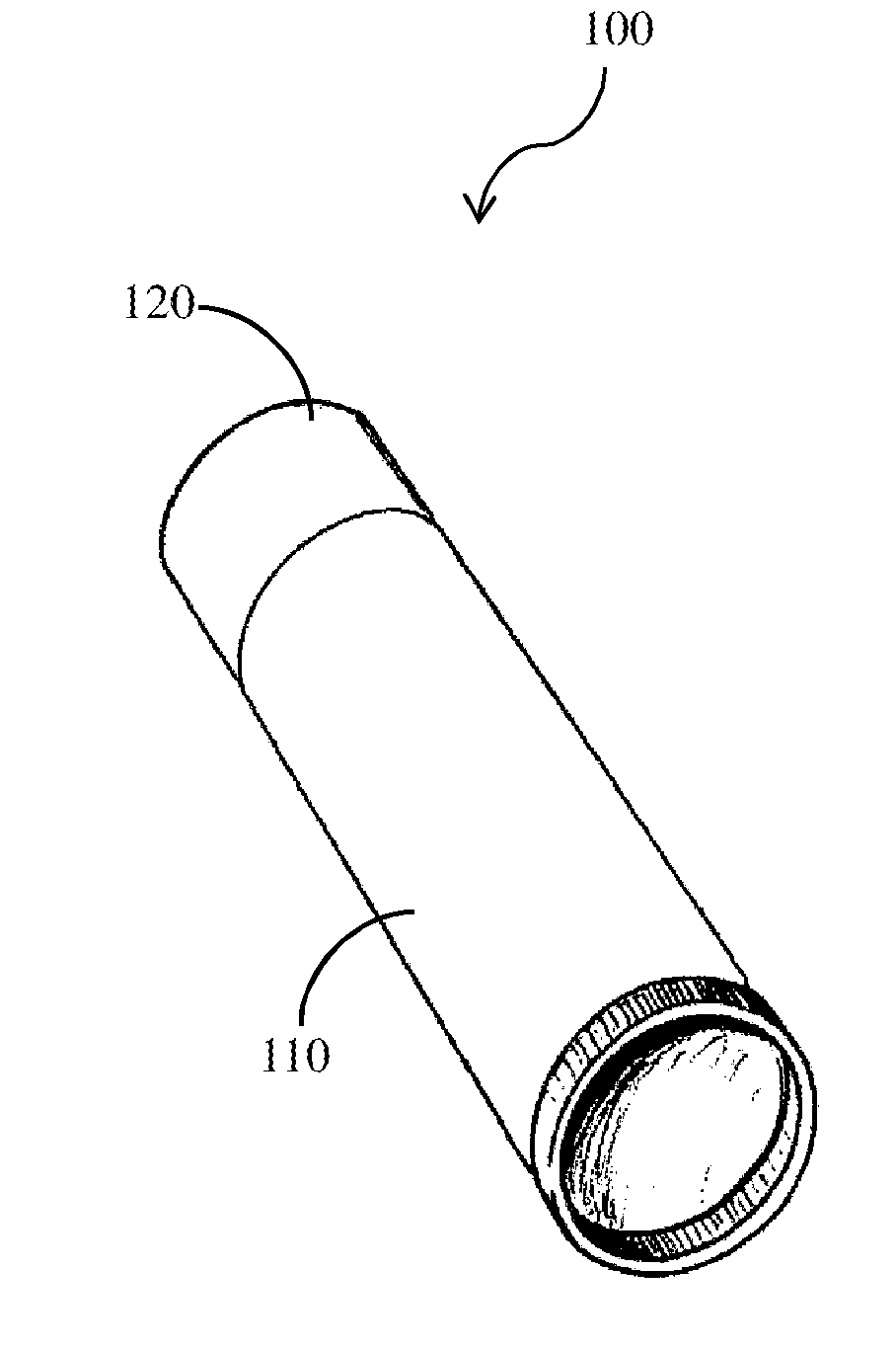
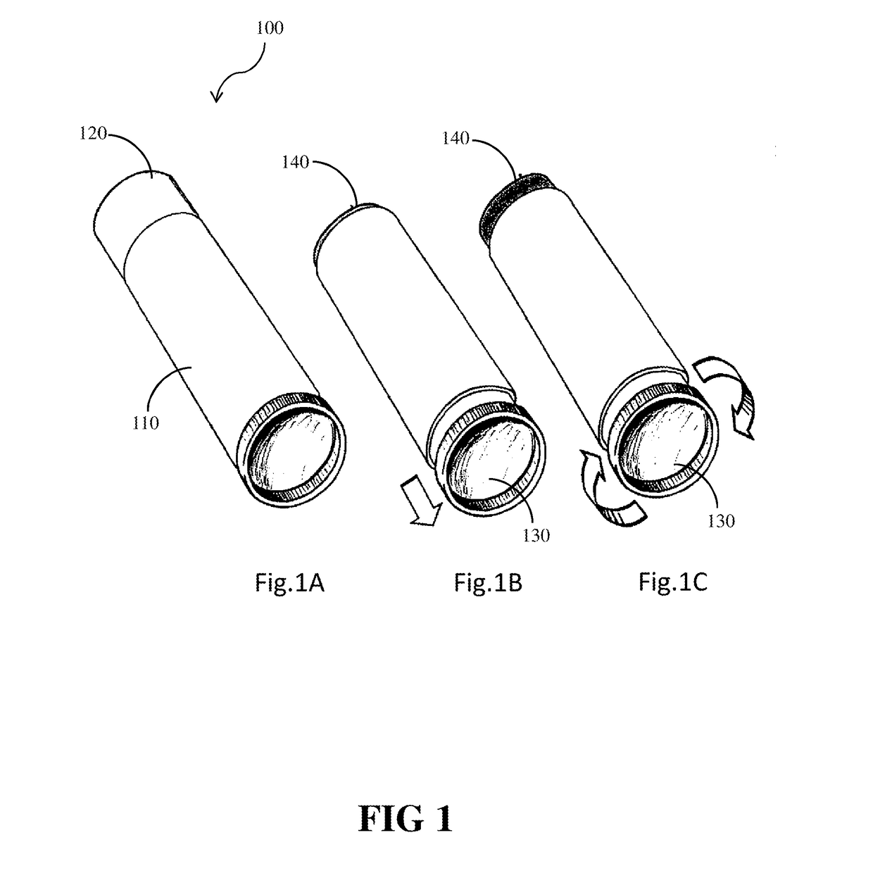
[0077]Formulation “90G” was made consisting entirely of Phospholipon 90G purified soy phosphatidylcholine, with a minimum purity of 94% phosphatidylcholine by weight. The yellowish, waxy solid material is supplied as small clumps. To form the material into a stick-form composition, it was repeatedly ground in a ceramic mortar and pestle that was heated to 40° C., then kneaded until solid, and then 4 gram aliquots were cold pressed into a 12 mm diameter cylinder.

example 3

[0078]Formulation “90G90H” was made by grinding together 6 grams of Phospholipon 90G and 3 grams of Phospholipon 90H (Lipoid Group, Köln, Germany). Phospholipon 90H is white powder purified soy derived phosphatidylcholine that is hydrogenated (fully saturated). The Phospholipon 90H was blended with the unsaturated natural phosphatidylcholine, and preheated to 60° C. to soften the hydrogenated form. The two materials were finely ground together in a ceramic mortar and pestle that was heated to 40° C. The mixture was kneaded until a smooth, solid, and cohesive waxy solid was created. Four gram aliquots were cold molded into 12 mm diameter cylindrical sticks.


PUM

PropertyMeasurementUnit
Fractionaaaaaaaaaa
Forceaaaaaaaaaa
Percent by massaaaaaaaaaa

Abstract

Methods for treating infection at the site of implantation of an orthopedic device in a human or animal subject. The methods include removing the orthopedic device, and implanting a replacement device. A surface of the replacement device is coated with an infection-inhibiting composition having a waxy matrix. The waxy matrix includes an infection-inhibiting material, such as a lipid, an antimicrobial agent, or a combination of a lipid and an antimicrobial agent.

Description

INTRODUCTION[0001]The present technology relates to orthopedic implants with an infection-inhibiting agent, methods for making orthopedic implants with an infection-inhibiting agent, and methods for treating infection at the site of implantation of an orthopedic device in a human or animal subject.[0002]Orthopedic implants are implantable medical devices used to replace, augment or repair bone, such as to replace diseased articulating joints (such as knees, hips and elbows), stabilize the skeleton where it has been destabilized by trauma (such as fractures), or to correct alignment. These implants are manufactured most commonly with plastics, polymers, ceramics, steel, stainless steel, metals and alloys.[0003]However, as foreign bodies, the surfaces of orthopedic devices implanted into the body provide a physical platform for bacteria to attach and grow. Due to the rapid growth rate and presence of virulence factors, bacteria are able to establish infections within days of the surgi...

Claims


Application Information

Patent Timeline
IPC IPC(8): A61L31/16A61L31/14A61K31/496A61B17/68A61K38/14A61K31/7036A61F2/30A61L31/08A61K31/65
CPCA61L31/16A61B2017/00889A61L31/146A61K31/496A61K31/65A61K38/14A61K31/7036A61F2/30771A61B17/68A61L2300/404A61L2300/406A61L2300/22A61F2310/0097A61F2002/3069A61F2002/3092A61F2002/30878A61F2002/30827A61L31/08A61L31/10A61F2/30767
Inventor TROXEL, KAREN S.
Owner BIOMET MFG CORP
Learn More