
Methods and systems for metal artifact reduction in spectral ct imaging

Active Publication Date: 2016-11-10
GENERAL ELECTRIC CO
View PDF0 Cites 33 Cited by
  • Summary
  • Abstract
  • Description
  • Claims
  • Application Information

AI Technical Summary

Benefits of technology

The patent describes a method for reducing metal artifacts in CT imaging by acquiring two sets of projections, detecting the location of metal in the first set, applying corrections to both sets of projections based on the location of metal, and displaying an image reconstructed from the corrected projections. This technique helps to improve the accuracy and reliability of CT imaging for diagnostic and therapeutic purposes.

Problems solved by technology

In some cases, the presence of metal (e.g., in the form of metal implants, dental fillings, and so on) may interfere with the x-ray attenuation, thereby causing metal artifacts in reconstructed images.
However, a simple application of known metal artifact reduction algorithms to dual energy CT leads to additional image artifacts.
Material decomposition utilizes data from both channels, and new artifacts arise due to the inconsistency of metal correction between the channels.

Method used


Image

  • Methods and systems for metal artifact reduction in spectral ct imaging
  • Methods and systems for metal artifact reduction in spectral ct imaging
  • Methods and systems for metal artifact reduction in spectral ct imaging

Examples


Embodiment Construction

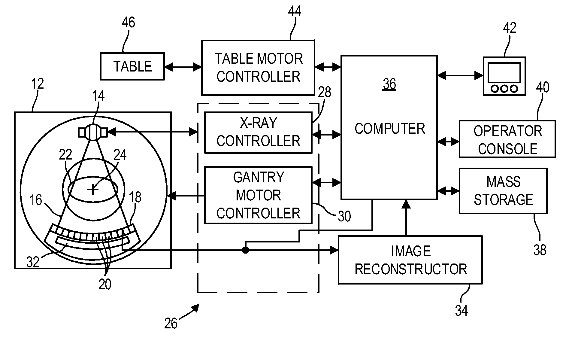
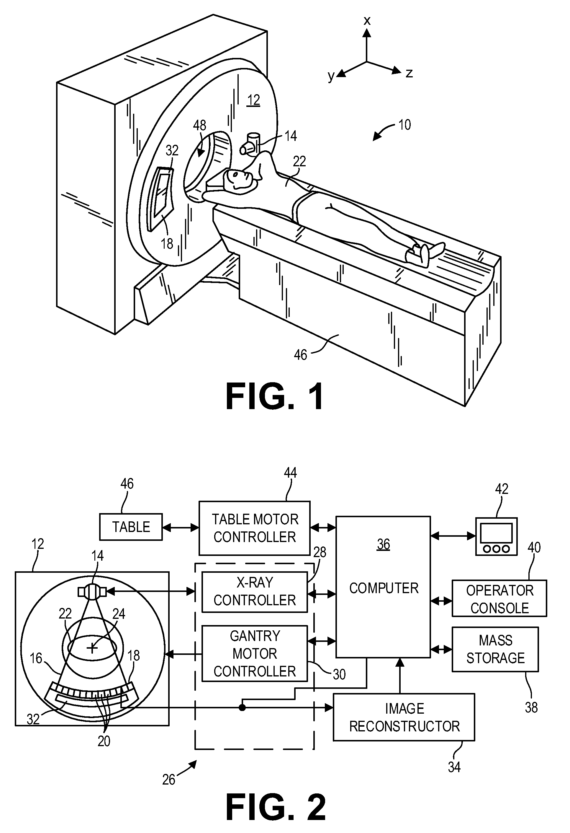
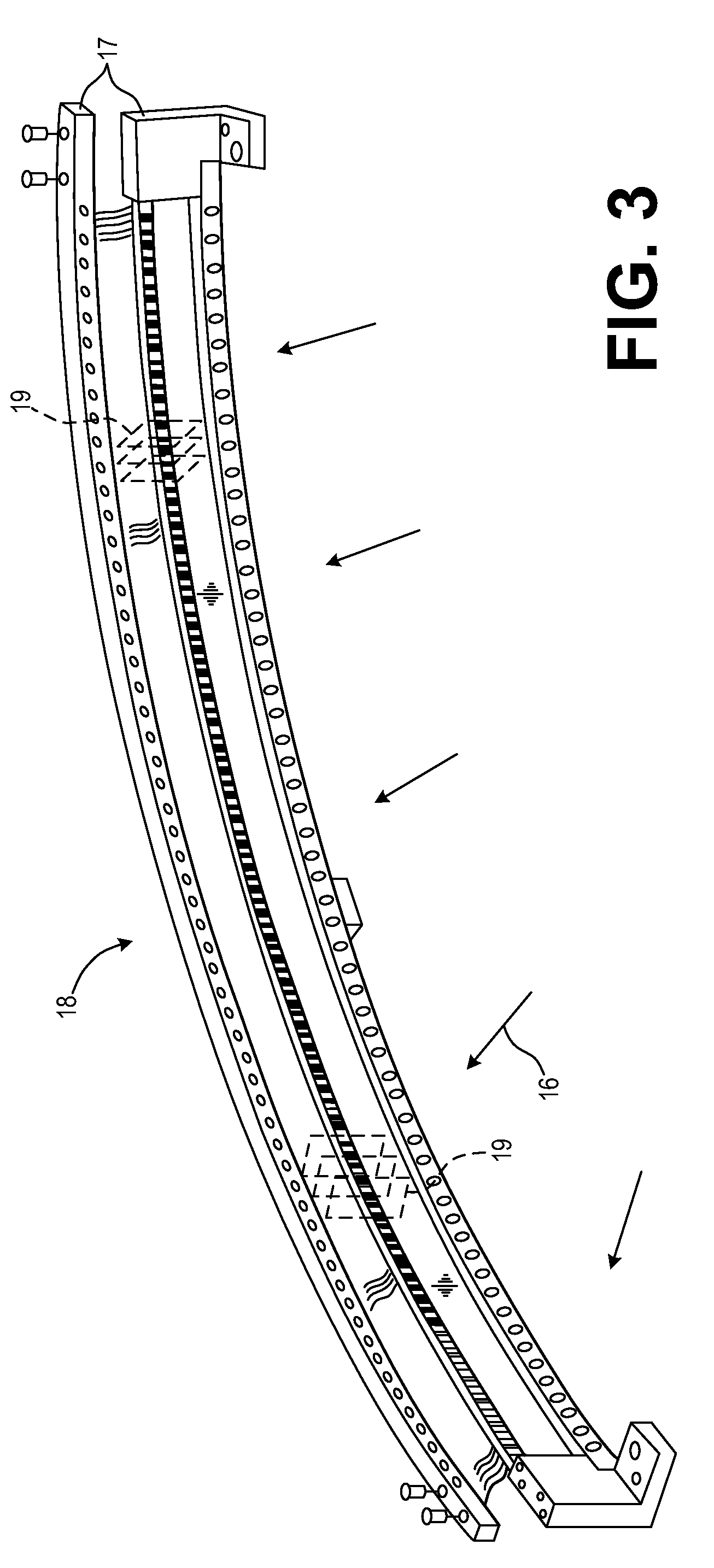
[0018]The following description relates to various embodiments of image reconstruction for dual energy spectral imaging. In particular, methods and systems for metal artifact reduction are disclosed. The operating environment of the present invention is described with respect to a sixty-four-slice computed tomography (CT) system, such as the CT imaging system shown in FIGS. 1-4. As described herein above, the presence of metal in an object being imaged (e.g., a patient, packages, etc.) may interfere with x-ray attenuation during CT imaging, thereby leading to metal artifacts in reconstructed images of the object. In dual or multi-energy CT imaging, multiple projection datasets may be acquired, where each projection dataset corresponds to a different acquisition energy. A method for dual or multi-energy imaging, such as the method depicted in FIG. 5, may include applying a metal artifact reduction algorithm to the multiple projection datasets, where the application of the metal artif...


PUM


Abstract

Various methods and systems for spectral computed tomography imaging are provided. In one embodiment, a method comprises acquiring a first projection dataset and a second projection dataset, detecting a location of metal in the first projection dataset, applying corrections to the first and second projection datasets based on the location of the metal, and displaying an image reconstructed from the corrected first and second projection datasets. In this way, metal artifacts may be substantially reduced in dual or multi-energy CT imaging.

Description

FIELD[0001]Embodiments of the subject matter disclosed herein relate to diagnostic imaging, and more particularly, to metal artifact reduction for dual energy spectral computed tomography (CT) imaging.BACKGROUND[0002]Dual or multi-energy spectral computed tomography (CT) systems can reveal the densities of different materials in an object and generate images acquired at multiple monochromatic x-ray energy levels. In the absence of object scatter, a system derives the behavior at a different energy based on a signal from two regions of photon energy in the spectrum: the low-energy and the high-energy portions of the incident x-ray spectrum. In a given energy region of medical CT, two physical processes dominate the x-ray attenuation: Compton scattering and the photoelectric effect. The detected signals from two energy regions provide sufficient information to resolve the energy dependence of the material being imaged. Detected signals from the two energy regions provide sufficient in...

Claims


Application Information

Patent Timeline
IPC IPC(8): A61B6/00G06T11/00A61B6/03
CPCA61B6/5258A61B6/482G06T2207/10081A61B6/032G06T11/008A61B6/461G06T11/005G06T2211/408
Inventor SEN SHARMA, KRITIGAO, HEWEIPAL, DEBASHISH
Owner GENERAL ELECTRIC CO
Learn More