
Systems and methods for feature detection in retinal images

Active Publication Date: 2016-10-06
DIGITAL DIAGNOSTICS INC
View PDF18 Cites 99 Cited by
  • Summary
  • Abstract
  • Description
  • Claims
  • Application Information

AI Technical Summary

Benefits of technology

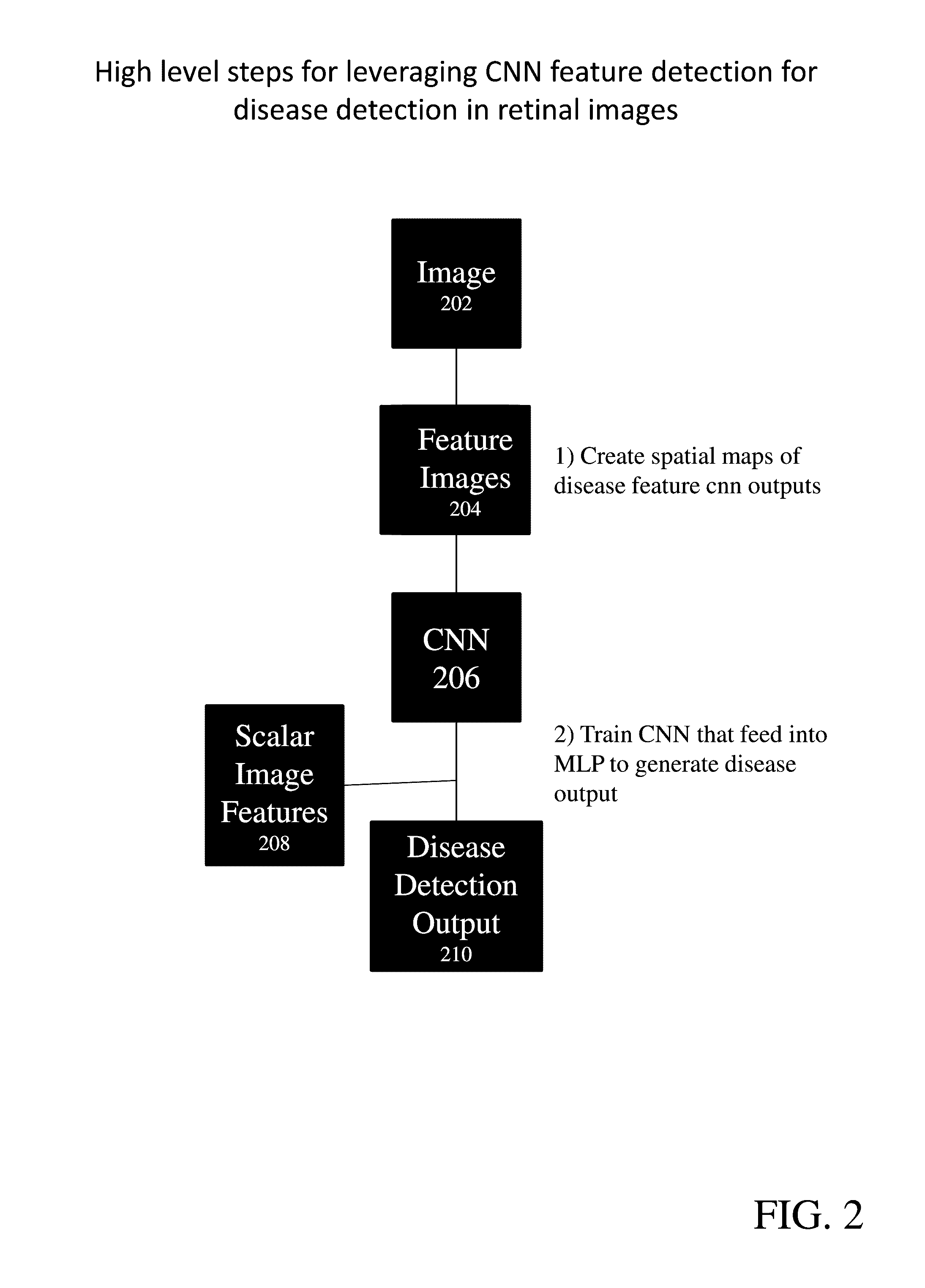
This patent presents a system and method for diagnosing disease in retinal images using a neural network. The system extracts image features from a retinal image, combines them with other features, and then inputs them into a trained neural network to identify disease. The system can also identify false positive and negative results and adjust the training dataset accordingly. The technical effect is a more accurate and reliable tool for diagnosing disease in retinal images.

Problems solved by technology

However, the straightforward use of NNs to classify entire retinal images or image subsets (patches) for these tasks, as is the art in other problems, may not lead to maximized performance in retinal images.
This is because the problem is underconstrained: there is a large variance in color, structure, and texture of the normal retina as well as a large diversity of objects, and a large variability in shapes, colors, textures, and other features of these objects, versus a relatively sparse number of retinal images with annotations at a sufficient level, that these machine learning algorithms require for optimal performance.
Medical images with annotations are sparse and expensive, because it both ultimately derives from patients so there are ethical concerns that prevent an unlimited number of images to be obtained, as well annotating these images which requires experts in that field, in contrast with other domains especially in computer vision where NNs are typically can be trained on unlimited number of images can be obtained and annotation can typically be done by any adult.
However, this still does not introduce sufficient ‘real’ variance, in both objects, as well as backgrounds.

Method used


Image

  • Systems and methods for feature detection in retinal images
  • Systems and methods for feature detection in retinal images
  • Systems and methods for feature detection in retinal images

Examples


examples

[0077]The following examples are put forth so as to provide those of ordinary skill in the art with a complete disclosure and description of certain examples of how the compounds, compositions, articles, devices and / or methods claimed herein are made and evaluated, and are intended to be purely exemplary of the invention and are not intended to limit the scope of what the inventors regard as their invention. However, those of skill in the art should, in light of the present disclosure, appreciate that many changes can be made in the specific embodiments which are disclosed and still obtain a like or similar result without departing from the spirit and scope of the invention.

[0078]FIG. 7 shows sample distribution with and without confounder training. As can be seen, the distribution of both the positive and negative samples improve (more negative samples pulled left and positive samples pushed right). At a threshold of 0.8, ˜33.5 samples marked as false positive would be expected. Af...


PUM


Abstract

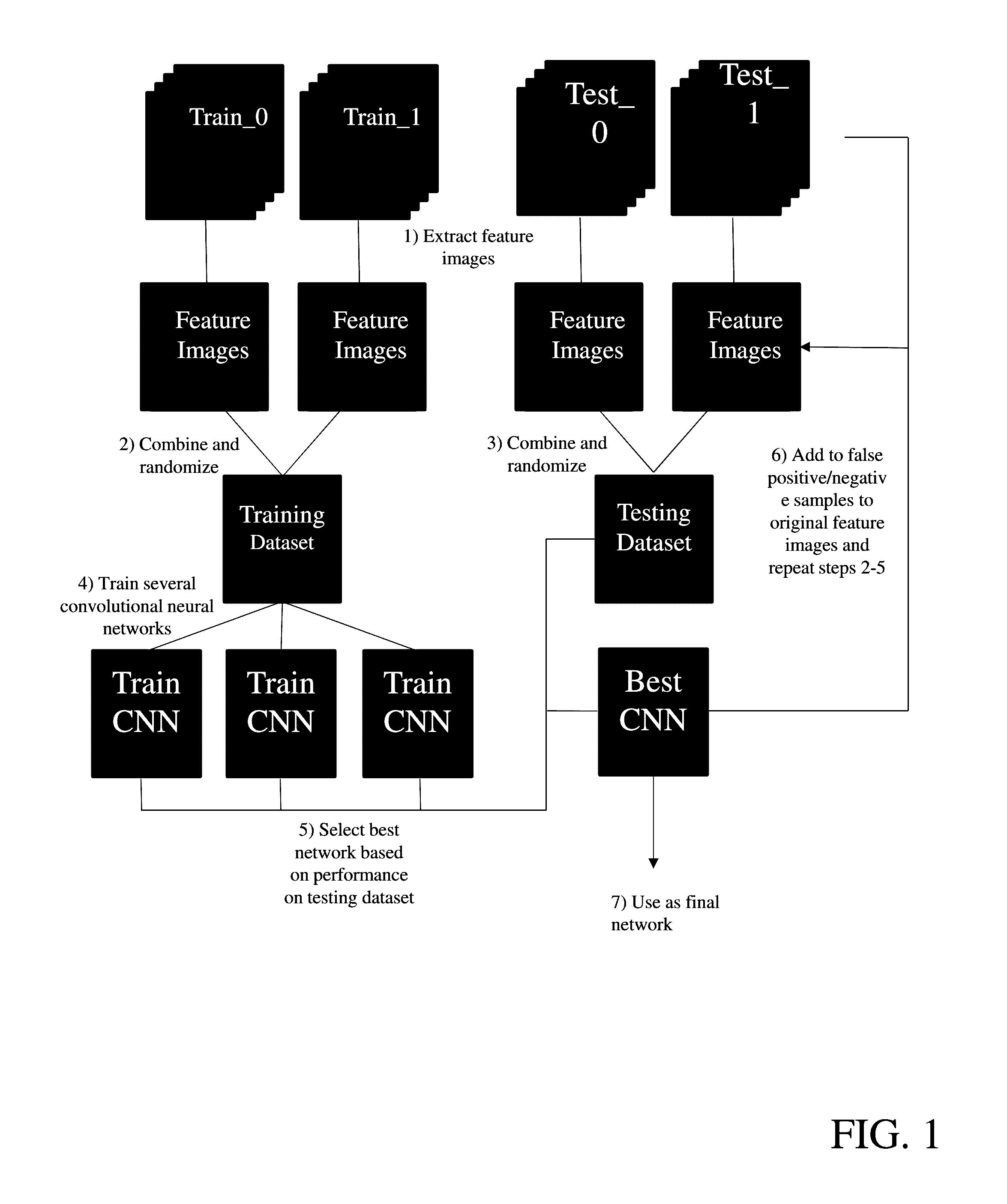
Provide are systems methods and devices for diagnosing disease in medical images. In certain aspects, disclosed is a method for training a neural network to detect features in a retinal image including the steps of: a) extracting one or more features images from a Train_0 set, a Test_0 set, a Train_1 set and a Test_1 set; b) combining and randomizing the feature images from Train_0 and Train_1 into a Training data set; c) combining and randomizing the feature images from Test_0 and Test_1 into a testing dataset; d) training a plurality of neural networks having different architectures using a subset of the training dataset while testing on a subset of the testing dataset; e) identifying the best neural network based on each of the plurality of neural networks performance on the testing data set; f) inputting images from Test_0, Train_1, Train_0 and Test_1 to the best neural network and identifying a limited number of false positives and false negative and adding the false positives and false negatives to the training dataset and testing dataset; and g) repeating steps d)-g) until an objective performance threshold is reached.

Description

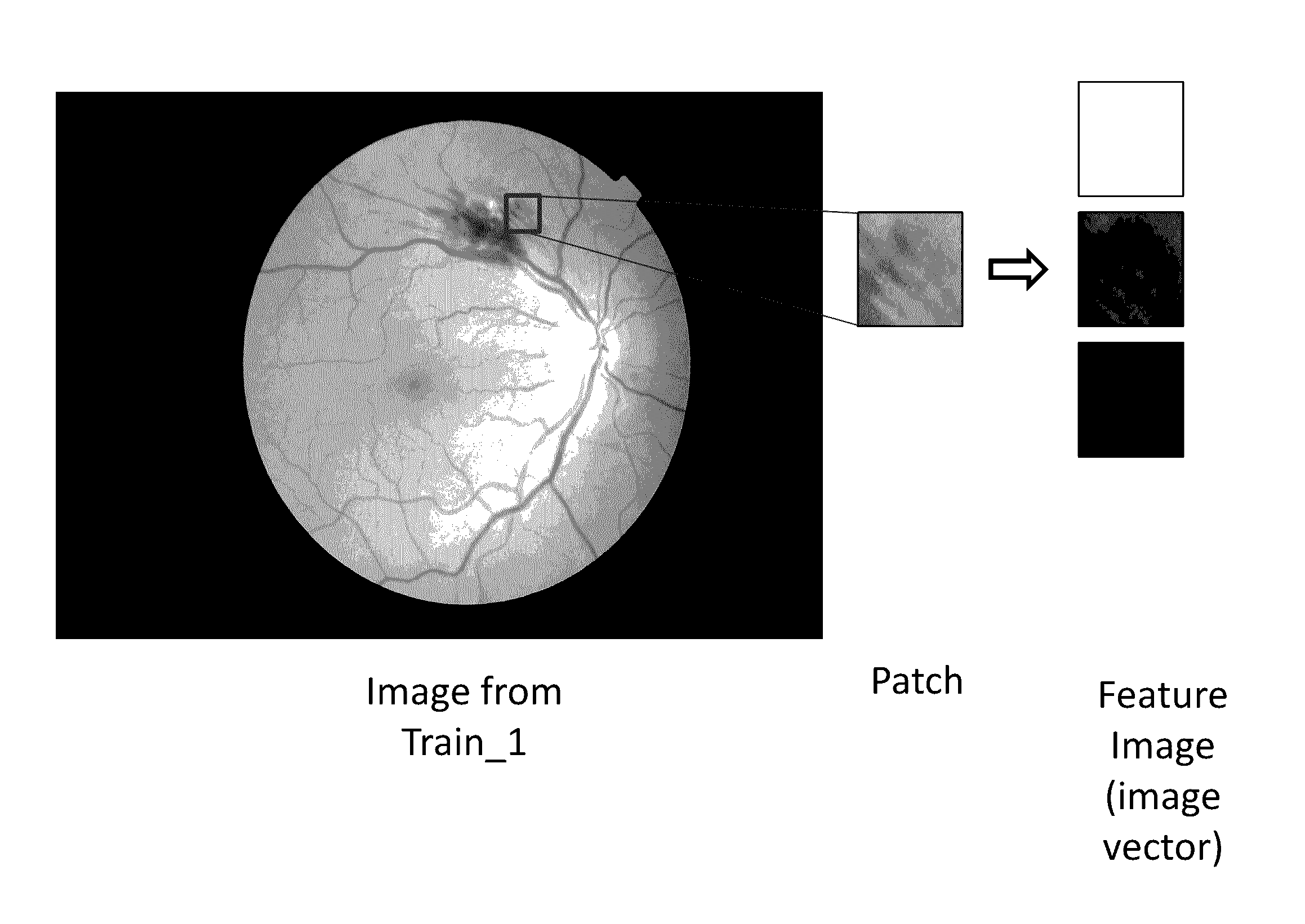
CROSS-REFERENCE TO RELATED APPLICATION(S)[0001]This application claims priority from U.S. Provisional Application No. 62 / 143,301, filed Apr. 6, 2015, which is hereby incorporated herein by reference in its entirety for all purposes.FIELD OF THE INVENTION[0002]Disclosed herein are systems and methods for retinal feature detection, learning, and classification for the purpose of retinal disease detection.BACKGROUND OF THE INVENTION[0003]Detecting, segmenting and classifying objects and other structures of interest in a background retinal, and even any other medical, image is crucial for detection and diagnosis of disease, its absence, as well as for biometrics. Deep neural networks (NNs), including Convolutional Neural Networks, as well as other types of multilevel neural networks, are an existing method for improved feature learning, classification, and detection. They have been applied to a wide range of different image types, including medical images). However, the straightforward ...

Claims


Application Information

Patent Timeline
IPC IPC(8): G06T7/00G06K9/46G06K9/62G06K9/66
CPCG06T7/0012G06K9/66G06K9/4604G06T2207/30041G06T2207/20084G06T2207/20081G06K9/6257G06N3/08G06V10/454G06V10/82G06N3/045G06F18/2148
Inventor NIEMEIJER, MEINDERTAMELON, RYANCLARIDE, WARRENTABRAMOFF, MICHAEL D.
Owner DIGITAL DIAGNOSTICS INC
Learn More