Systems and Method for Computation and Visualization of Segmentation Uncertainty in Medical Images

a technology of segmentation uncertainty and computation method, applied in the field of medical image uncertainty, can solve problems such as difficulty in correctly delineating the boundary of the anatomy, noisy parts of the model boundary, blurry parts,

Active Publication Date: 2016-09-15
SIEMENS HEALTHCARE GMBH
View PDF2 Cites 12 Cited by
  • Summary
  • Abstract
  • Description
  • Claims
  • Application Information

AI Technical Summary

Benefits of technology

The patent text describes a system and method for computing the uncertainty of a target anatomical object using medical imaging data. The uncertainty is estimated at each of a plurality of vertices on a surface model of the object. The uncertainty may be visualized on the model, and can be refined based on the range of points along the surface normal for each vertice. The system can also use marginal space learning to segment the object and determine its boundaries. The measurement of the patient based on the surface model and uncertainty can include a range of values, a mean, and a standard deviation with a confidence interval.

Problems solved by technology

However, due to image noise and ill-defined boundaries caused by neighboring tissues with a similar imaging response, parts of the model boundary can be noisy, blurry, or have signal dropout.
It is often challenging to correctly delineate the boundary of the anatomy in these situations.

Method used


Image

  • Systems and Method for Computation and Visualization of Segmentation Uncertainty in Medical Images
  • Systems and Method for Computation and Visualization of Segmentation Uncertainty in Medical Images
  • Systems and Method for Computation and Visualization of Segmentation Uncertainty in Medical Images

Examples


Embodiment Construction

[0020]The present invention generally relates to computation and visualization of segmentation uncertainty in medical images. Embodiments of the present invention are described herein to give a visual understanding of methods for computing and visualizing segmentation uncertainty in medical images. A digital image is often composed of digital representations of one or more objects (or shapes). The digital representation of an object is often described herein in terms of identifying and manipulating the objects. Such manipulations are virtual manipulations accomplished in the memory or other circuitry / hardware of a computer system. Accordingly, is to be understood that embodiments of the present invention may be performed within a computer system using data stored within the computer system.

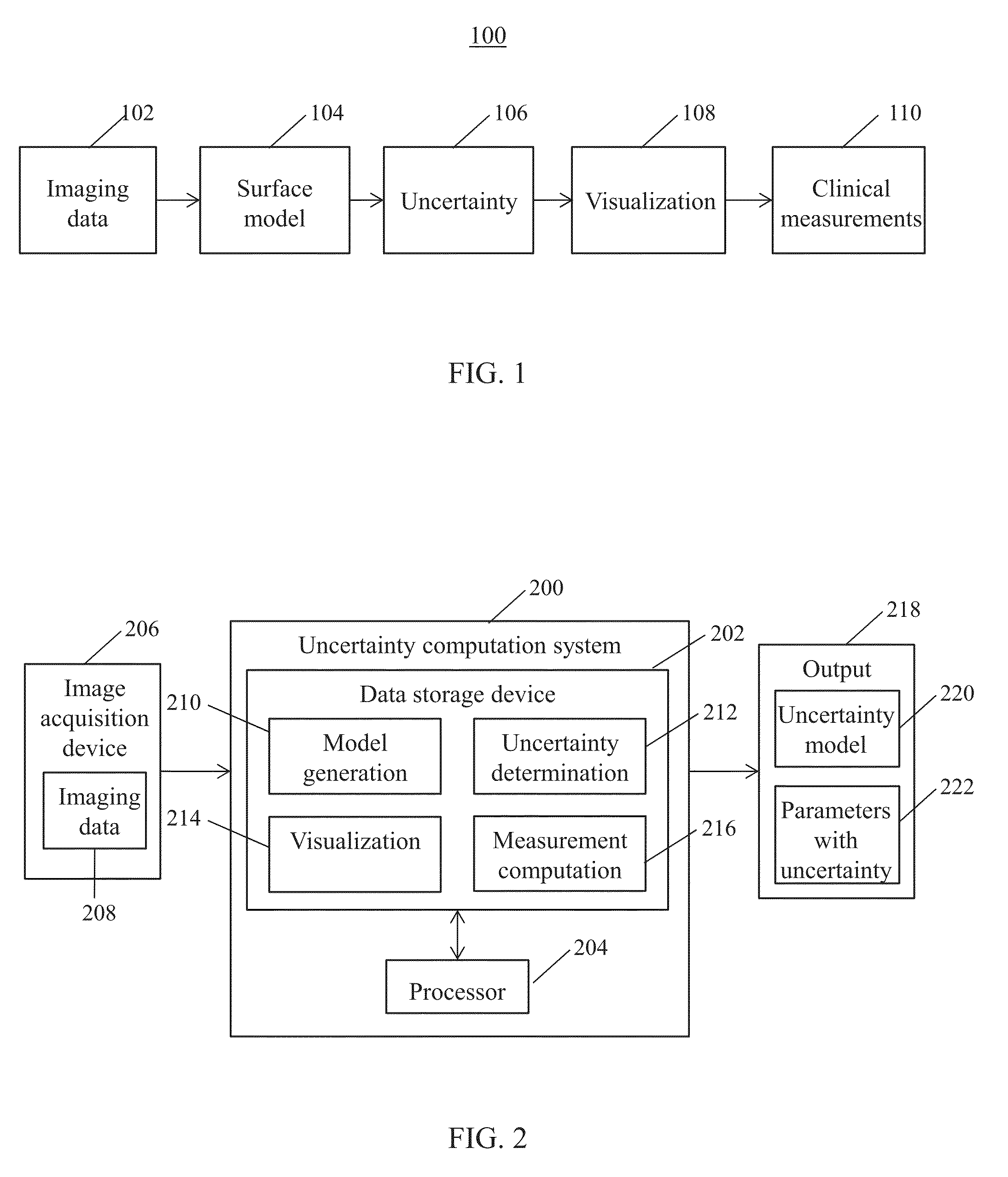
[0021]FIG. 1 shows a high-level framework 100 for computing segmentation uncertainty, in accordance with one or more embodiments. Framework 100 generates a surface model of an anatomical area of...


PUM


Abstract

Systems and methods for computing uncertainty include generating a surface model of a target anatomical object from medical imaging data of a patient. Uncertainty is estimated at each of a plurality of vertices of the surface model. The uncertainty estimated at each of the plurality of vertices is visualized on the surface model.

Description

BACKGROUND OF THE INVENTION[0001]The present invention relates generally to uncertainty in medical images and more particularly to computation and visualization of segmentation uncertainty in medical images.[0002]Many clinical applications nowadays rely on geometrical models for quantification of physiology and function. In a standard setting, these models are generated from two-dimension (2D), three-dimensional (3D), and 3D plus time (3D+t) medical images. Based on the estimated models, clinically relevant measurements are computed and used for diagnosis and intervention planning. For example, during transcatheter aortic valve implantation (TAVI) planning, a 3D computed tomography (CT) or transesophageal echocardiogram (TEE) image is acquired. Based on the 3D image, a patient specific surface model of the aortic valve can be extracted and distance measurements for implant sizing are derived. However, due to image noise and ill-defined boundaries caused by neighboring tissues with a...

Claims


Application Information

Patent Timeline
Patent Type & Authority Applications(United States)
IPC IPC(8): G06T7/00G06F19/00G16H30/40
CPCG06T7/0081G06T7/0012G06F19/321G06T7/0097G06T7/0085G06T2207/20192G06T2207/30004G06T2207/10072G06T2207/20221G06T2210/12G06T2210/41G06T2207/30048G06T2207/30056G06V2201/031G06F18/285G06T2200/04G06T2207/10081G06T2207/20081G06T2207/30061G06T19/00G06T7/174G06T7/62G06T7/11G16H30/40
Inventor GRBIC, SASAMANSI, TOMMASOVOIGT, INGMARGEORGESCU, BOGDANFLORIN, CHARLES HENRICOMANICIU, DORIN
Owner SIEMENS HEALTHCARE GMBH
Why Eureka
Try Eureka