
Portable medical device and method for quantitative retinal image analysis through a smartphone

a medical device and smartphone technology, applied in image analysis, medical science, diagnostics, etc., can solve the problems of limited clinical use of fundoscopy, retinal image acquisition relying on expensive and non-portable devices, and analysis of retinal images, so as to achieve the effect of inexpensive hardware and extremely portabl

Active Publication Date: 2015-11-12
UNIV DELLA CALABRIA
View PDF0 Cites 12 Cited by
  • Summary
  • Abstract
  • Description
  • Claims
  • Application Information

AI Technical Summary

Benefits of technology

The present invention is a system that allows for the capture and analysis of high-resolution images of the retina using a smartphone-adapted lens and a software app. This system uses cloud-based technologies for computation and can provide quantitative indices of retinal damage that are useful in both clinical and research settings.

Problems solved by technology

However, full exploitation of fundoscopy in clinical settings is currently quite limited due to two main reasons:acquisition of retinal images mostly relies upon expensive and non-portable devices;analysis of retinal images is limited to qualitative, naked-eye assessment of retinal abnormalities in routine clinical practice.
However, these solutions succeeded in improving either portability or analysis, neglecting to integrate the innovations and thus to obtain an inexpensive, portable technology that addresses both needs.
To date, a few innovations have been developed in order to acquire retinal images through a portable device, such as a smartphone; however, it is worth noting that none of them is coupled with a software that performs quantitative analyses of the images.
It is worth stating that none of the abovementioned technologies perform quantitative image analysis of the retina.
However, this technology is available only for workstations.
While a number of portable instruments were developed for acquisition of retinal images and softwares were developed for image analysis, none of the available tools integrate, within a single, inexpensive, portable device (such as a smartphone), both improved acquisition and quantitation of the retinal image.
As a major limitation, iExaminer does not perform any quantitative analysis of the retinal images.
Furthermore, the adapter system is relatively cumbersome.

Method used


Image

  • Portable medical device and method for quantitative retinal image analysis through a smartphone
  • Portable medical device and method for quantitative retinal image analysis through a smartphone
  • Portable medical device and method for quantitative retinal image analysis through a smartphone

Examples


Embodiment Construction

[0018]The present invention consists in an optical device for the acquisition of high-resolution retinal images through a smartphone equipped with camera and a light source, where the acquired images will be processed by the smartphone app in order to obtain quantitative indices of retinal damage.

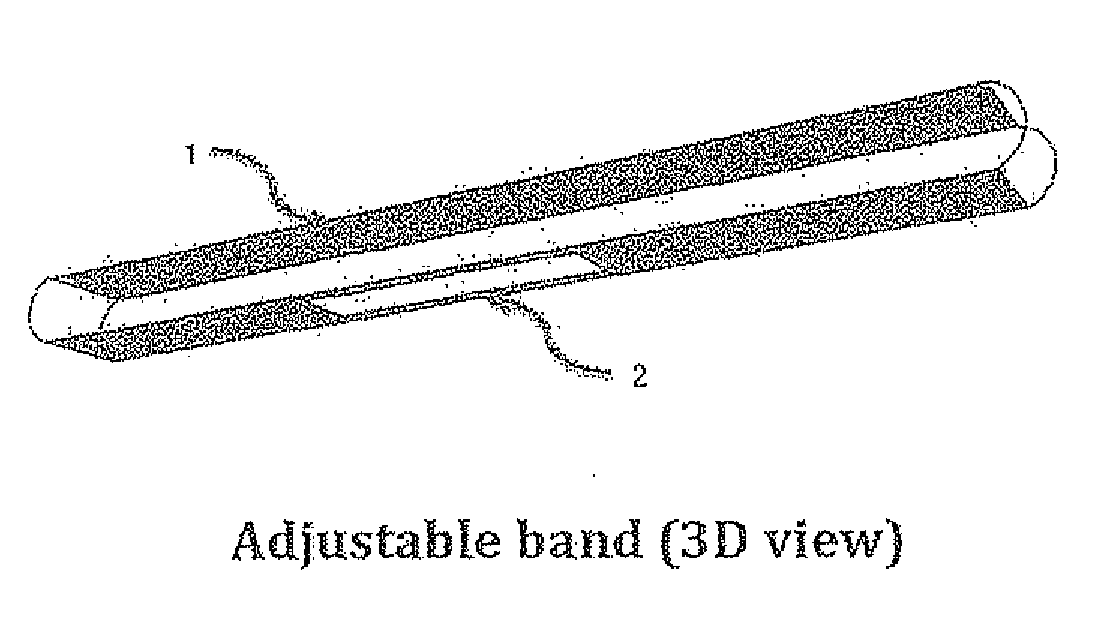
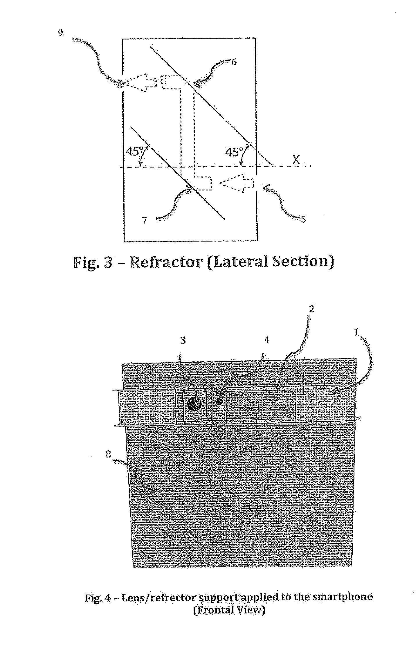
[0019]The optical device includes:[0020]smartphone (8), FIG. 4;[0021]adjustable band (I), FIG. 1, with a built-in sliding track (2), FIG. 1;[0022]lens (3), FIG. 2;[0023]refractor (4), FIG. 2.

[0024]The adjustable band (1), FIG. 1, is mounted on the smartphone (8), FIG. 4 to allow the positioning of the lens (3), FIG. 2 and refractor (4), FIG. 2, in front of the smartphone's camera and flash light, respectively. The adjustable band (1), FIG. 1, mounted on the smartphone body (8), FIG. 4 is represented in FIG. 4.

[0025]The lens (3), FIG. 2—and the refractor (4), FIG. 2, are mounted on the sliding track (2), FIG. 1, of the adjustable band (1), FIG. 1, next to each other (3,4) FIG. 2. By means of...


PUM


Abstract

The present invention essentially consists of an integrated system that allows acquisition and quantitative analysis of high-resolution images of the retina. The integrated system includes an optical device and a software app. The optical device is a smartphone-adapted lens for acquisition of high-resolution images of the retina. The software app is a smartphone app for quantitative analysis of the retinal images acquired through the hardware component of the device. The computation is carried out by means of cloud-based technologies. Quantitative indices of retinal damage are obtained. These indices are proven for being relevant for both clinical and research purposes.

Description

TECHNICAL FIELD[0001]The innovation herewith presented refers to the technical field of portable medical devices. Specifically, the innovation consists in a portable medical device that allows acquisition and analysis of retinal images through a mobile device, such as a smartphone.STATE OF THE ART[0002]Examination of the retina through fundoscopy is a key medical procedure for diagnosis and monitoring of a number of pathological conditions, including ophthalmologic (e.g. glaucoma and macular degeneration), endocrinologic (e.g. diabetes), cardiovascular (e.g. hypertension) and neurologic disorders (e.g. brain tumors, head trauma, cerebrovascular diseases, dementia and multiple sclerosis). However, full exploitation of fundoscopy in clinical settings is currently quite limited due to two main reasons:[0003]acquisition of retinal images mostly relies upon expensive and non-portable devices;[0004]analysis of retinal images is limited to qualitative, naked-eye assessment of retinal abnor...

Claims


Application Information

Patent Timeline
IPC IPC(8): A61B3/14A61B3/00
CPCA61B3/0008A61B3/14A61B3/12G06T7/0012G06T2207/30041G06T2207/10024G06T2207/20044G06T7/11G06T7/155G06T7/194
Inventor STAMILE, CLAUDIOUMETON, RENATOCAVALLARI, MICHELECALIMERI, FRANCESCOORZI, FRANCESCO
Owner UNIV DELLA CALABRIA
Learn More