
Method and analysis device for microscopic examination of a tissue section or cell smear

a tissue section or cell smear technology, applied in the field of immunological and/or histochemical examination devices, can solve problems such as inability to charge, inability to perform the analysis, and considerable difficulty

Inactive Publication Date: 2015-07-30
EUROIMMUN MEDIZINISCHE LABORDIAGNOSTIKA
View PDF1 Cites 12 Cited by
  • Summary
  • Abstract
  • Description
  • Claims
  • Application Information

AI Technical Summary

Benefits of technology

The invention is about a new system for processing samples, particularly in tumour diagnosis. The system uses small, flexible substrate supports that can hold multiple samples, such as biochips, and can be easily combined and separated for further processing and analysis. The substrate supports can be made of plastic instead of glass, which makes them more flexible and easier to work with. The system also includes a holder that can securely hold the substrate supports and samples for processing and evaluation. The holder has a surface that can hold the substrate supports, and the substrate supports can be easily released and connected again. The system also includes a groove or trough to prevent liquids from mixing together during processing. Overall, the system allows for more efficient and flexible processing of samples, reducing the need for manual handling and the risk of contamination.

Problems solved by technology

However, it is only with some considerable difficulty that tissue sections from different areas of a piece of tissue can be arranged on a slide and examined simultaneously.
The problem of the described technical solution is that it is only with some considerable difficulty that it is possible for tissue sections from different areas of a piece of tissue to be arranged on a slide as precisely as would be desirable for rapid and effective examination.
However, a flexible arrangement of sample fragments, which allows substrate supports to be charged in the required manner, such that the charging is modified, for example depending on a planned work step in the laboratory, is not possible with the described technical solution.

Method used


Image

  • Method and analysis device for microscopic examination of a tissue section or cell smear
  • Method and analysis device for microscopic examination of a tissue section or cell smear
  • Method and analysis device for microscopic examination of a tissue section or cell smear

Examples


Embodiment Construction

[0026]The invention is explained in more detail below with reference to the figures, without limiting the general concept of the invention. In the figures:

[0027]FIG. 1 shows a substrate support with samples of biological material arranged thereon;

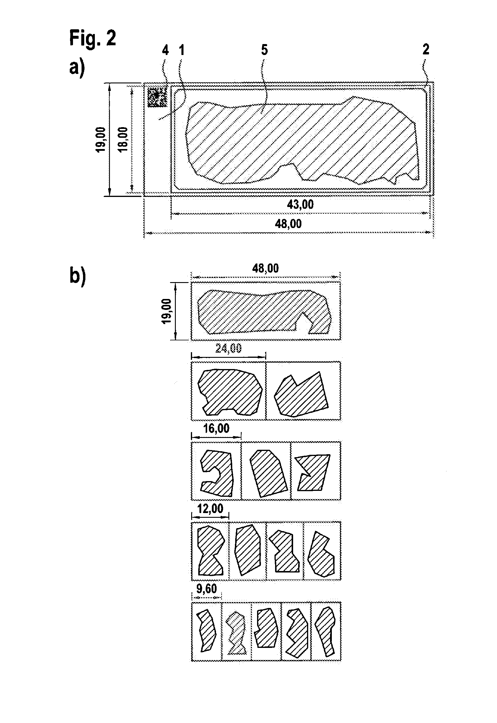
[0028]FIG. 2 shows various arrangements of samples of biological material on substrate supports;

[0029]FIG. 3 shows different views of a holder in the form of a diagnostic frame with charged substrate supports;

[0030]FIG. 4 shows different ways of charging a holder in the form of a diagnostic frame;

[0031]FIG. 5 shows a plan view of a tablet with a plurality of holders;

[0032]FIG. 6 shows different views of a holder in the form of an incubation holder with groove-shaped liquid receptacles;

[0033]FIG. 7 shows a schematic view of the incubation of substrate supports with samples of biological material, which are arranged in groove-shaped liquid receptacles of an incubation holder;

[0034]FIG. 8 shows different views of a holder in the form of an inc...


PUM


Abstract

The invention relates to a method and a device for microscopically examining patient samples consisting of tissue or cells. Especially in the field of tumor diagnostics, typically the entire samples or selected sections thereof that are of special interest for test purposes are initially placed on substrate supports one by one or in groups. Said substrate supports are then placed individually or together with other substrate supports in holders in which the substrate supports are subjected to a defined sequence of process steps. According to the invention, the substrate supports that contain the patient samples are securely immobilized in the holders and can be rearranged differently for subsequent laboratory operations. For each laboratory operation, the patient samples thus form combined groups that are processed in parallel or are moved jointly.

Description

TECHNICAL FIELD[0001]The invention relates to a device and a method for immunological and / or histochemical examination of patient samples in the form of tissue sections and / or cells. After they have been fixed on a substrate support and then stained with the aid of a suitable reactant, the respective patient samples undergo a microscopic examination for diagnosis.PRIOR ART[0002]In the field of laboratory diagnostics, various analysis methods are known in which patient samples in the form of tissue sections or cells are each initially stained, and the stained structures are then examined to establish a diagnosis. Particularly in the area of histology or histopathology, micrometre-thin, stained tissue sections are produced and assessed on a microscope. The sample material in histological work includes in particular surgical specimens, sample excisions, and tissue removed by means of biopsies, the main aim of the examination of tissue sections stained in this way being the reliable det...

Claims


Application Information

Patent Timeline
IPC IPC(8): G01N1/31G02B21/34G01N33/50B01L9/00G01N33/569
CPCG01N1/312B01L9/523G02B21/34G01N33/5005G01N33/56966G01N35/00029B01L2300/0822G01N2035/00138G01N2035/00158G01N2035/00752
Inventor STOCKER, WINFRIEDROTTMANN, NORBERT
Owner EUROIMMUN MEDIZINISCHE LABORDIAGNOSTIKA
Learn More