Method of examining mammary gland disease

a mammary gland and disease technology, applied in the field of mammary gland disease detection, can solve the problems of difficult to discover and medically treat latent mastitis, low clinical expression of latent mastitis, and spread of latent mastitis among cow hosts without being noticed, so as to achieve easy and quick identification, easy and quick implementation, and the effect of prolonging the duration of infection

Active Publication Date: 2015-07-09
TOHOKU UNIV
View PDF3 Cites 1 Cited by
  • Summary
  • Abstract
  • Description
  • Claims
  • Application Information

AI Technical Summary

Benefits of technology

[0064]According to the method of the present invention, a site in a subject that is infected by a mammary gland disease can be identified easily and quickly if compared with any known comparable methods as a result of using as indicator the expression level of cyclophilin A in milk or in mammary gland tissue obtained from the subject. The method of the present invention is particularly effective for identifying an udder or an udder quarter that is in initial stages of mastitis. Additionally, since the method of the present invention is a non-invasive method, it is possibly applicable to prevention of mastitis.
[0065]Since the method of the present invention can be put to practice easily and quickly, the method enables onsite measurements in dairy farms. If it is possible to determine mastitis in an udder or an udder quarter in early stages of the disease, milking can be suspended early, a treatment can be started early with the use of a small amount of antibiotic agent, and the duration of infection can be curtailed and milking can be resumed early so that treatment expenses can be suppressed and the economic loss of the dairy farmer feeding the cow due to the reduction of income caused by a reduced amount of milk obtained by milking can be minimized.
[0066]Cows are most liable to be infected by mastitis in the postpartum period at and near the time point of a month after

Problems solved by technology

The frequency of clinical expressions of latent mastitis is relatively low, it is difficult to discover and medically treat latent mastitis.
The problem is that the latent mastitis can spread among hosts of cows without being noticed.
However, when it comes to mastitis caused by Staphylococcus aureus, part of Staphylococcus aureus is resista

Method used


Image

  • Method of examining mammary gland disease
  • Method of examining mammary gland disease
  • Method of examining mammary gland disease

Examples


examples

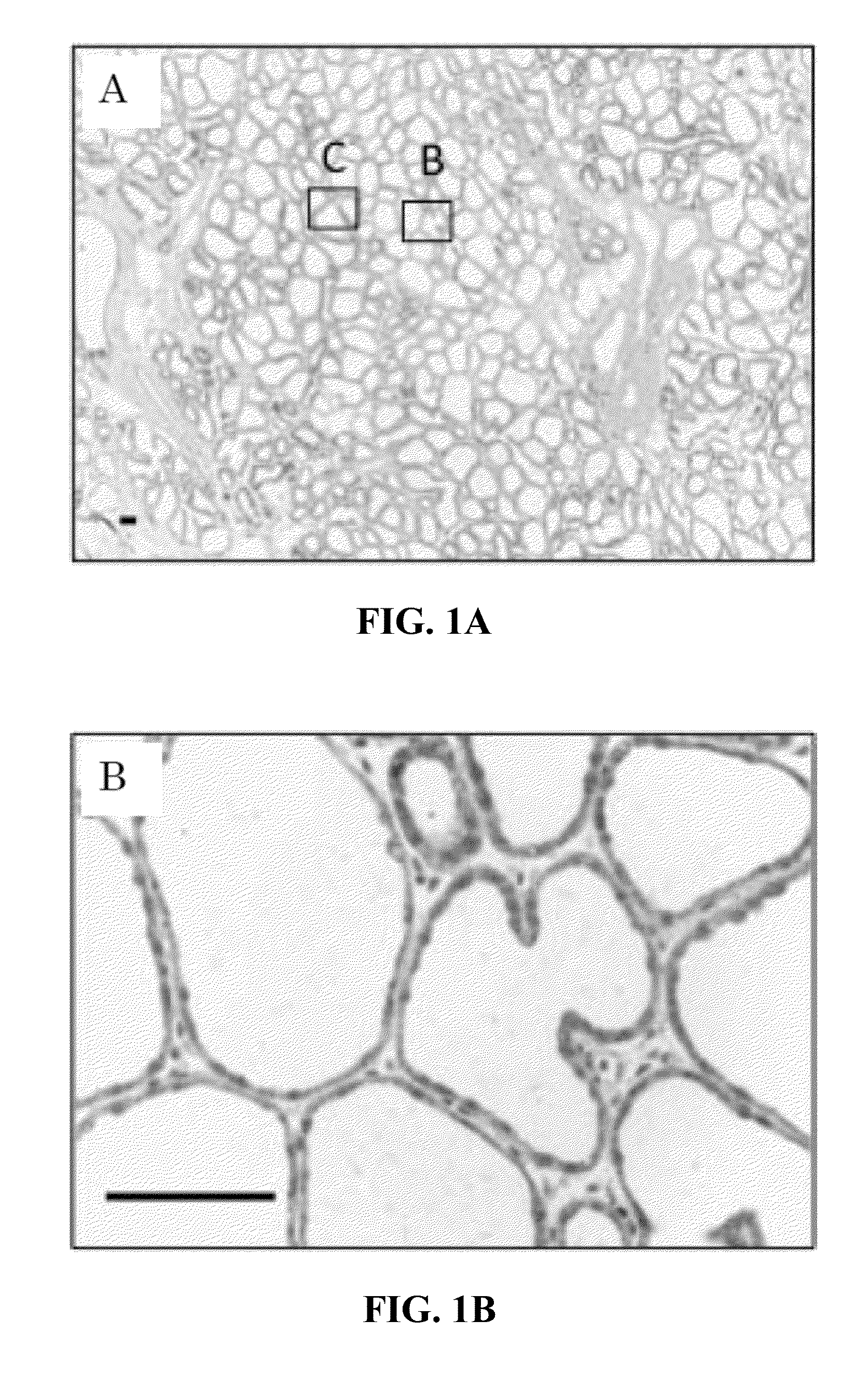
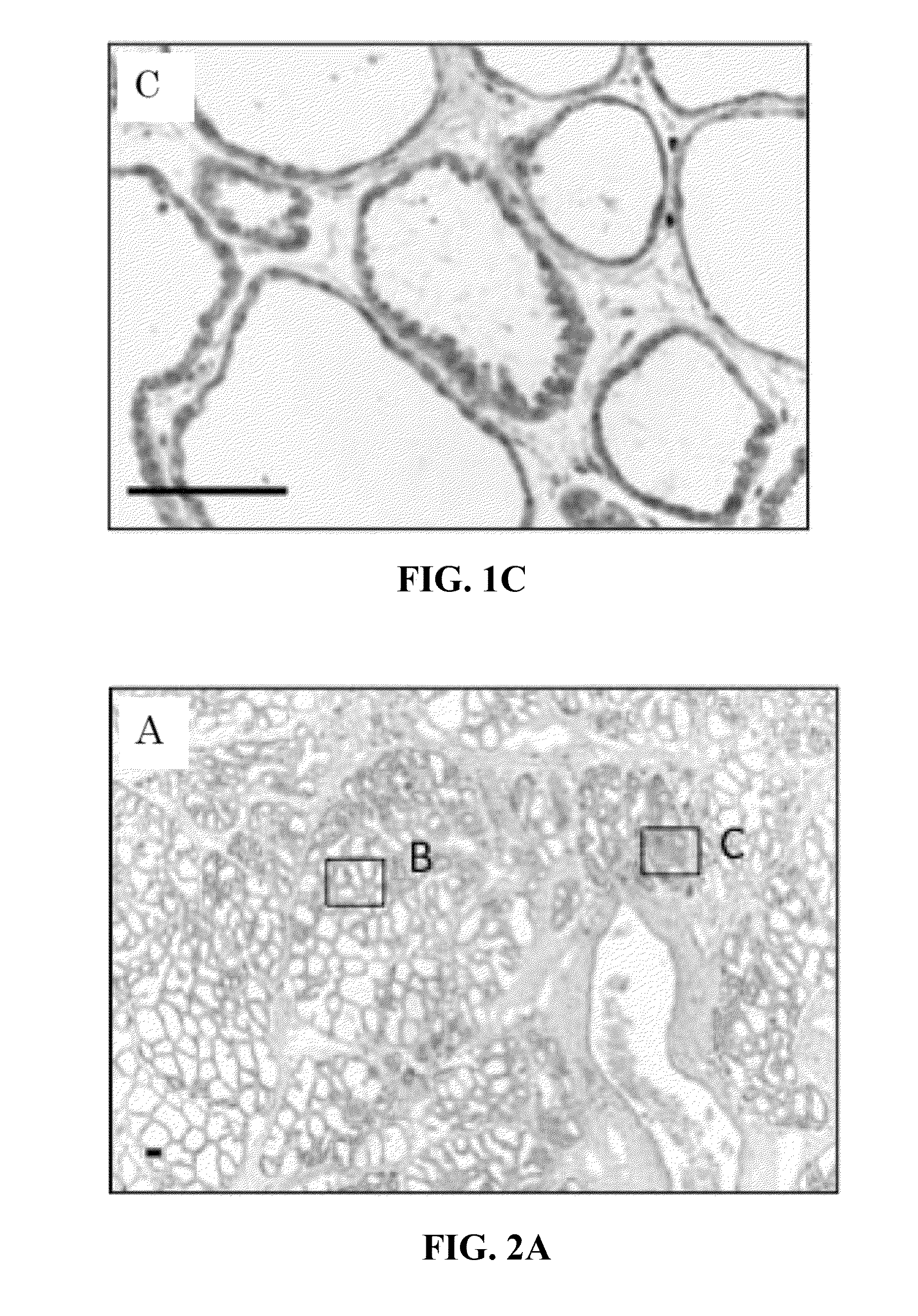
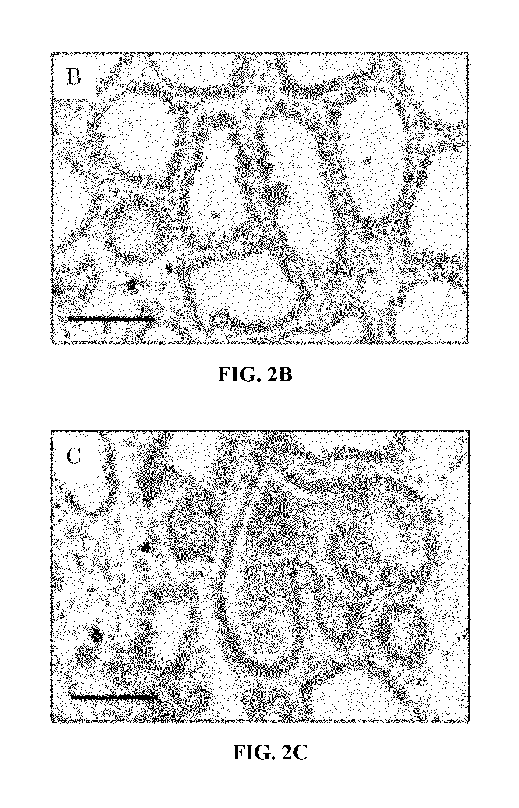
1st Localization of Cyclophilin A in Mammary Gland Tissue

1. Materials and Methods

(1) Samples

[0174]The mammary gland tissues collected from the udders where the onset of experimental mastitis or latent mastitis was observable out of lactating Holstein cows of three different experimental districts indicated as A through C below were selected and used as mammary gland tissues developing mastitis. The mammary gland tissues were collected immediately after slaughtering the cows and were immobilized in a single night at 4° C. by quickly using PLP fixative solution or phosphoric acid buffer formalin fixative solution. After the fixation, the tissues were immersed sequentially in 70% ethanol, in 80% ethanol, in 90% ethanol and in 95% ethanol for 12 hours each and then in 100% ethanol for 24 hours so as to be dehydrated stepwise. After the dehydration, the tissues were immersed in each of toluene and paraffin for 6 hours and then embedded in paraffin. Additionally, the mammary gland tissues...


PUM

PropertyMeasurementUnit
Levelaaaaaaaaaa

Abstract

The object of the present invention is to provide an early mastitis detection method that can detect mastitis easily and quickly when compared to the prior methods and also a biomarker to be used with the method.
The object can be solved by a method of examining a mammary gland disease by using the level of cyclophilin A in a mammary gland or in milk as indicator. More specifically, the object can be a method of examining a mammary gland disease comprising steps (1) and (2) listed below;
  • (1) a step of detecting cyclophilin A in the milk collected from an udder or an udder quarter of a subject and thereby determining the cyclophilin A level in the milk; and
  • (2) a step of determining the onset of a mammary gland disease or the possibility of onset of a mammary gland disease in the udder or the udder quarter of the subject on the basis of the cyclophilin A level in the milk.

Description

[0001]The present application claims the benefit of priority of Japanese Patent Application No. 2012-156163, filed on Jul. 12, 2012, the entire contents of which are incorporated herein by reference.TECHNICAL FIELD[0002]The present invention relates to the detection method of mammary gland disease such as mastitis in the udder or in one or more of quarter parts of udder.BACKGROUND ART[0003]The cow udder is divided into the left and the right by a thick medial suspensory ligament, and also into the front and the rear by thin membranes that operate as partition wall to constitute independent quarters. The cow udder contains a large number of mammary alveoli, and a single layer of mammary epithelial cells is arranged at the mammary gland side of each mammary alveolus. Mammary epithelial cells of a healthy cow are tightly connected by inter-cellular adhesion molecules such as tight junctions. Mammary epithelial cells form a layer, which functions as barrier for physically separating the...

Claims


Application Information

Patent Timeline
IPC IPC(8): G01N33/573C07K16/40
CPCG01N33/573G01N2800/365G01N2333/99C07K16/40G01N33/6893
Inventor ASO, HISASHIKITAZAWA, HARUKIWATANABE, KOICHIOWADA, SHUICHIWATANABE, HITOSHINAGASAWA, YUYASOMEYA, SHUNSUKEHORIKOSHI, YORIKOITAYA, NANAMI
Owner TOHOKU UNIV
Why Eureka
Try Eureka