Camera Arrangement and Image Processing Method for Quantifying Tissue Structure and Degeneration

Inactive Publication Date: 2015-02-19
BIOOPTICO
View PDF31 Cites 6 Cited by
  • Summary
  • Abstract
  • Description
  • Claims
  • Application Information

AI Technical Summary

Benefits of technology

The invention describes a way to use optics to enhance and visualize the structure of cartilage, and a algorithm to quantify how much it's degenerating. This can be done using existing cameras in arthroscopic procedures. The technique involves getting data, converting it to light, filtering it to enhance the structure, and measuring how much it's fluctuating. The results are then displayed in a more easily accessible way. Overall, the invention provides a way to automatically enhance and quantify cartilage structure and degeneration.

Problems solved by technology

The fibrillation of cartilage consequently leads to cartilage softening and, with time, deeper cartilage defects.

Method used


Image

  • Camera Arrangement and Image Processing Method for Quantifying Tissue Structure and Degeneration
  • Camera Arrangement and Image Processing Method for Quantifying Tissue Structure and Degeneration
  • Camera Arrangement and Image Processing Method for Quantifying Tissue Structure and Degeneration

Examples


Embodiment Construction

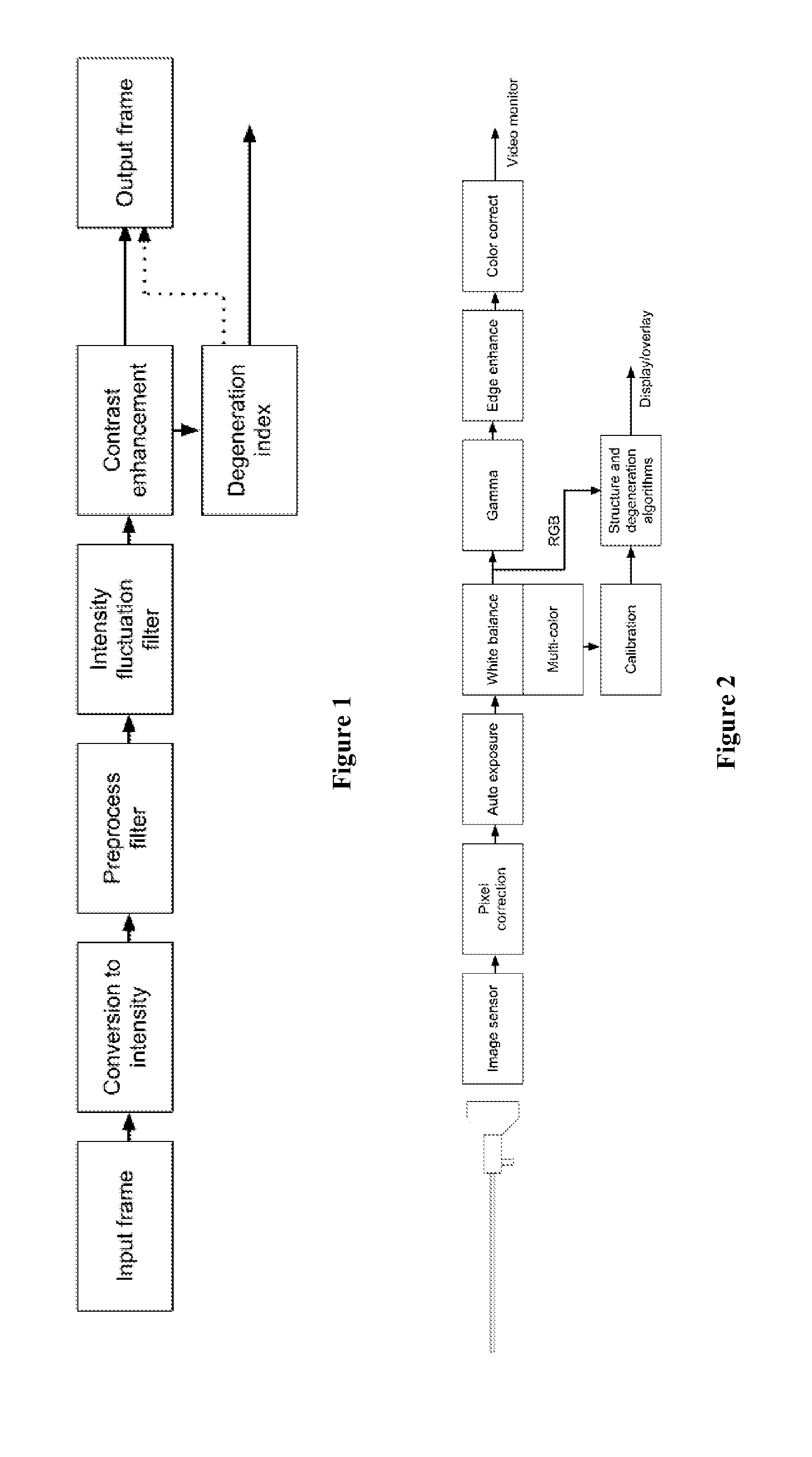
[0018]The main steps of the tissue structure enhancement method according to the invention can be summarized as: Obtaining input data, conversion to intensity data, preprocess filtering, intensity fluctuation filtering, contrast enhancement and output frame presentation (FIG. 1). These different steps are described below, together with a description of the DI calculation.

Obtaining Input Data

[0019]Circuitry and processing within an endoscopic video camera are well suited to perform algorithm calculations and graphically present the result as an enhancement to the live arthroscopic image. For purposes of displaying the best endoscopic image on the surgical monitor, the raw red, green and blue (RGB) signals collected by the camera and endoscope are, in endoscopic cameras used today, modified by both linear and non-linear transformations. For example, edge enhancement, color correction and gamma correction. Such transformations may affect the quality of algorithm calculations. On the ot...


PUM


Abstract

Methods and arrangements for detecting osteoarthritis (OA) relate to image processing for enhancing, visualizing and quantifying the fibrillation structure of cartilage using endoscopes. A structure enhancement method comprises obtaining input data, conversion to intensity data, preprocess filtering, intensity fluctuation filtering and contrast enhancement. The degeneration is quantified by a degeneration index (DI) algorithm, applied to the structure enhanced image. Results are then compiled in an output frame presentation.

Description

TECHNICAL FIELD[0001]The present invention relates to methods and arrangements for detecting osteoarthritis (OA). In particular the present invention relates to image processing for enhancing, visualizing and quantifying the fibrillation structure of cartilage using endoscopes.BACKGROUND OF THE INVENTION[0002]Osteoarthritis is the most common type of joint disease, affecting over 20 million individuals in the United States. The condition involves degeneration of articular cartilage and subchondral bone in joints. Typical symptoms include joint pain, stiffness and locking. The processes leading to loss of cartilage are still not fully known, but include a variety of hereditary, metabolic and mechanical factors.[0003]Cartilage is a type of connective tissue, made up of cells (chondrocytes) embedded in a matrix, strengthened with collagen fibers. One of the earliest signs of OA is fibrillation of the collagen structure, seen both as roughening of the cartilage surface and as deeper str...

Claims


Application Information

Patent Timeline
IPC IPC(8): A61B1/317A61B1/04G06T5/00
CPCA61B1/317G06T5/009A61B1/04G06T2207/30024G06T2207/10024G06T2207/20172G06T2207/10016A61B5/4514G06T7/0012G06T2207/10068G06T2207/30008G06V20/695A61B1/000094A61B1/000095G06T5/90G06T5/92
Inventor JOHANSSON, ANDERSOBERG, KE P.SUNDQVIST, TOMMYPERSSON, FREDRIK
Owner BIOOPTICO
Why Eureka
Try Eureka