
Photoacoustic tracking and registration in interventional ultrasound

a technology of ultrasound and photoacoustic tracking, applied in the field of intraoperative registration and tracking systems, can solve the problems of adding complexity, limiting the application of conventional methods, and affecting the effect of the patient's recovery

Active Publication Date: 2015-01-29
THE JOHN HOPKINS UNIV SCHOOL OF MEDICINE
View PDF9 Cites 65 Cited by
  • Summary
  • Abstract
  • Description
  • Claims
  • Application Information

AI Technical Summary

Benefits of technology

The current invention is an intraoperative registration and tracking system that uses optical and ultrasound imaging to locate and track tissue during surgery. The system includes an optical source, an optical imaging system, an ultrasound imaging system, and a registration system that combines the optical and ultrasound images. This allows for a more accurate and precise mapping of tissue layers and enables surgeons to better target and remove abnormal tissue while minimizing damage to surrounding healthy tissue.

Problems solved by technology

One significant factor limiting the use of IOUS is the necessity for the surgeon to relate the information in the ultrasound images to preoperative information such as CT or MRI images and to what he or she is seeing in a laparoscopic video monitor or in direct viewing.
There are important limitations with the conventional approaches.
However, these approaches have serious limitations.
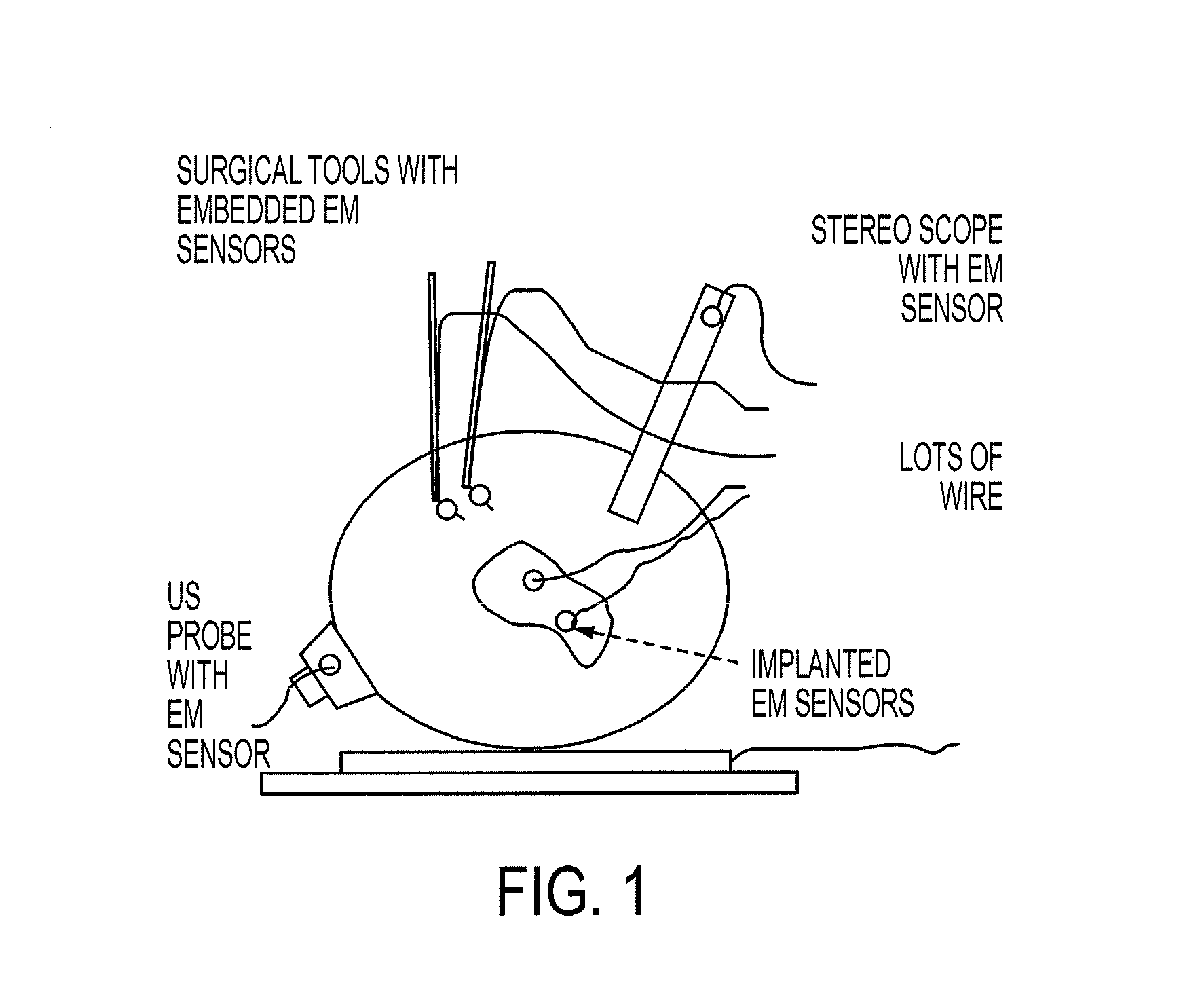
Navigational trackers typically track sensors or markers relative to a separate base station placed somewhere close to the surgical environment, thus adding complexity.
Electromagnetic systems require wires between the sensors and base unit, thus complicating sterility, are subject to field distortions, and may not work well in the presence of metal.
Accuracy in estimating tool tip position is limited by tool shaft bending and the effects of angle estimation error if markers are placed at a distance from the tip, and it is often impractical to embed sensors near the tips of small tools such as needles or the tines of ablation probes.
In addition, the estimation of IOUS-to-camera or IOUS-to-tool transformations necessarily requires an indirect calculation based on multiple tracking targets and is subject to error buildup.
Furthermore, calibration of US imaging probes to tracking devices is tedious.

Method used


Image

  • Photoacoustic tracking and registration in interventional ultrasound
  • Photoacoustic tracking and registration in interventional ultrasound
  • Photoacoustic tracking and registration in interventional ultrasound

Examples


example 1

[0053]Although the interventional PA techniques according to some embodiments of the current invention are can be broadly applicable, in the current example, we use laparoscopic partial nephrectomies (LPN) and RF ablation of tumors in solid organs such as the liver and kidney. For the LPN application a typical workflow would be as follows: Preoperative CT would be used to plan the surgical resection. Intraoperatively, the surgeon would position the kidney so that the tumor is close to the surface facing the surgeon and a 3DUS probe would be placed on the opposite side of the kidney in a position where the tumor, surrounding tissue, and organ surface is visible in the ultrasound. PA-to-video registration would be performed continuously using a system according to an embodiment of the current invention. 3DUS-to-CT registration would be performed and overlay images would be generated on ultrasound and video images, showing the segmented tumor and resection plan. Using this information,...

example 2

REFERENCES FOR EXAMPLE 2

[0168]1. Wang Y., Butner S., and Darzi A.: The developing market for medical robotics. Proceedings of the IEEE 94(9), 1763-1771, September (2006).[0169]2. Taylor, R., Lavallee, S., Burdea, G., and Mosges, R.: Computer integrated surgery. MIT Press Cambridge, Mass. (1996).[0170]3. Stolka P. J., Keil M., Sakas G., McVeigh E., Allaf M. E., Taylor R. H., and Boctor E. M.: A 3D-elastography-guided system for laparoscopic partial nephrectomies. in Medical Imaging 2010: Visualization, Image-Guided Procedures, and Modeling, San Diego, February 13-18, 76251I, 76251I-12 (2010)[0171]4. Boctor E., Viswanathan A., Choti M., Taylor R., Fichtinger G., and Hager G.: A Novel Closed Form Solution for Ultrasound Calibration. in International Symposium on Biomedical Imaging, Arlington, 527-530 (2004)[0172]5. Poon T. and Rohling R.: Comparison of calibration methods for spatial tracking of a 3-D ultrasound probe. Ultrasound in Medicine and Biology 31(8), 1095-1108, August (2005)[...

example 3

[0185]In this example we refine and evaluate the registration required to bring the preoperative prostate MRI model into the da Vinci visualization system. To achieve this goal we perform the following three tasks to be executed in the following order:

[0186]Task 1:

[0187]3DUS B-mode and PA-mode reconstruction

[0188]Rationale:

[0189]Volumetric intraoperative ultrasound is used to fuse the pre-operative MRI model to the surgical scene. In general, 3DUS data can be acquired using two different approaches. One approach is to utilize a 2D ultrasound array to directly provide 3DUS B-mode data. Unfortunately, these 2D arrays are not widely available and to the best of our knowledge, there is no 2D TRUS array. Alternatively, there are a number of mechanical probes that provide 3DUS data by wobbling a 1D array, but these are relatively slow and need customization to synchronize with a PA imaging system. The second approach is to track a conventional 1D TRUS probe using mechanical, optical or el...


PUM


Abstract

An intraoperative registration and tracking system includes an optical source configured to illuminate tissue intraoperatively with electromagnetic radiation at a substantially localized spot so as to provide a photoacoustic source at the substantially localize spot, an optical imaging system configured to form an optical image of at least a portion of the tissue and to detect and determine a position of the substantially localized spot in the optical image, an ultrasound imaging system configured to form an ultrasound image of at least a portion of the tissue and to detect and determine a position of the substantially localized spot in the ultrasound image, and a registration system configured to determine a coordinate transformation that registers the optical image with the ultrasound image based at least partially on a correspondence of the spot in the optical image with the spot in the ultrasound image.

Description

CROSS-REFERENCE OF RELATED APPLICATION[0001]This application claims priority to U.S. Provisional Application No. 61 / 608,910, filed Mar. 9, 2012, the entire contents of which are hereby incorporated by reference.BACKGROUND[0002]1. Field of Invention[0003]The field of the currently claimed embodiments of this invention relates to intraoperative registration and tracking systems, and more particularly to intraoperative registration and tracking systems that use photoacoustic tracking and registration in interventional ultrasound.[0004]2. Discussion of Related Art[0005]Intraoperative ultrasound (IOUS) imaging can be extremely useful in surgery and other interventional procedures. Ultrasound (US) systems provide real time 2D and 3D views into the patient's anatomy with relatively unobtrusive hardware and without the use of ionizing radiation. An increasing variety of transcutaneous, laparoscopic, and catheter-based probes including both 2D and 3D arrays are available from multiple vendor...

Claims


Application Information

Patent Timeline
IPC IPC(8): A61B8/08A61B5/00A61B8/14A61B8/00
CPCA61B8/5261A61B8/4416A61B5/0084A61B5/0095A61B8/14A61B8/0841A61B6/032A61B6/5247A61B8/483A61B8/4254A61B8/445A61B2090/373A61B2034/2063A61B2090/3945A61B34/20A61B2090/364A61B90/361A61B2090/306A61B90/37A61B2090/371A61B2034/2065A61B2090/378A61B2090/365A61B2090/3937
Inventor BOCTOR, EMAD M.TAYLOR, RUSSELL H.KANG, JIN U.
Owner THE JOHN HOPKINS UNIV SCHOOL OF MEDICINE
Learn More