
Spinal canal access and probe positioning, devices and methods

a technology for locating probes and accessing the spinal canal, which is applied in the field of spinal canal accessing and probe positioning, devices and methods, and can solve problems such as cerebrospinal fluid leakage, csf leakage, and faulty positioning or placement of probes or catheters

Active Publication Date: 2015-01-22
MEDLINE IND LP
View PDF46 Cites 21 Cited by
  • Summary
  • Abstract
  • Description
  • Claims
  • Application Information

AI Technical Summary

Benefits of technology

This solution significantly reduces the risk of complications by providing immediate and accurate feedback on needle placement, reducing procedure time, and minimizing the chances of accidental dural puncture or incorrect catheter placement, thereby enhancing the efficiency and safety of spinal canal access procedures.

Problems solved by technology

If the needle is put in too far, it will pass through the epidural space and puncture a thin layer of tissue (the dura), entering the subarachnoid space and causing a cerebrospinal fluid (CSF) leak.
Unfortunately, complications due to faulty positioning or placement of the probe or catheter are not uncommon during epidural procedures.
One of the most frequent complications occurs when the epidural needle is accidentally inserted past the epidural space and through the dura, resulting in a cerebrospinal fluid (CSF) leak.
These headaches are often severe and associated with nausea and vomiting, vision and hearing changes, low back pain, dizziness, and cranial nerve palsies.
Additionally, if left untreated, the headaches can predispose to subdural hematoma and possibly death.
Another common error during epidural anesthesia occurs when a catheter is introduced in an area other than the epidural space, like the surrounding muscles.
This error happens because, due to tissue structure differences, these areas can give a false “loss of resistance” upon epidural needle entry.
Unfortunately, it is difficult and time consuming to identify misplaced catheters.
The drug will not take effect if the catheter is not in the epidural space, and since peak effect of correctly delivered drug can take up to 20 minutes, verification by this method can be time consuming.
Such a delay can be impractical for a patient in severe pain, and may in fact be dangerous for a woman in need of an urgent caesarean section.
Both problems, puncturing the dura and putting the catheter in the wrong place, result because the “loss-of-resistance” technique is simply not particularly sensitive.
Further, there is a lack of a suitable alternative that does not involve impractical complexity.
Infants commonly require lumbar puncture as a part of the routine workup for fever without a source, as they have a much higher risk of meningitis than adults and do not reliably show external signs of meningitis, like a stiff neck.
Unfortunately, the “pop” is not as noticeable in some adult patients as well as in infants generally.
The CSF may take tens of seconds to make its way out the end of the needle, and the physician may have to reposition the needle multiple times before seeing CSF return through the needle, making the procedure very time consuming and uncomfortable for the patient.
If the physician advances the spinal needle too far, the needle can damage the blood vessels at the distal side of the CSF space and cause what is known as a “bloody tap” that contaminates the CSF sample and frequently leads to additional procedures.
Unfortunately, nearly 20% of pediatric lumbar punctures result in a “bloody tap”, in part because the physicians do not have a good indication of when the needle has entered the correct space.
It can be difficult to perform this measurement for a number of reasons, including the following: potential for attachment and removal of the apparatus and patient movement to dislodge the needle; relatively long time (e.g., several minutes) to acquire a reading; potential for inaccuracy from air bubbles; loss of CSF increasing post-dural puncture headache frequency.

Method used


Image

  • Spinal canal access and probe positioning, devices and methods
  • Spinal canal access and probe positioning, devices and methods
  • Spinal canal access and probe positioning, devices and methods

Examples


Embodiment Construction

[0035]The present invention provides methods and structures for detecting and / or facilitating positioning of a probe in the spinal canal of a patient, including probe positioning, monitoring and the like during a spinal canal access procedure, such as a lumbar puncture or epidural access procedure.

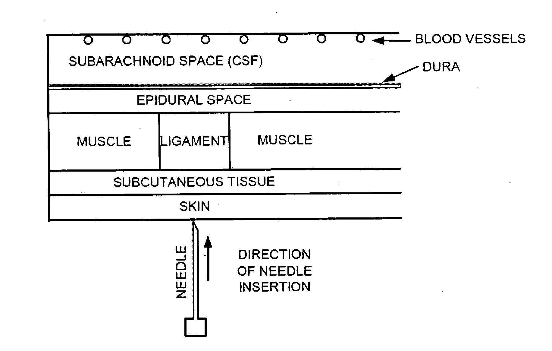
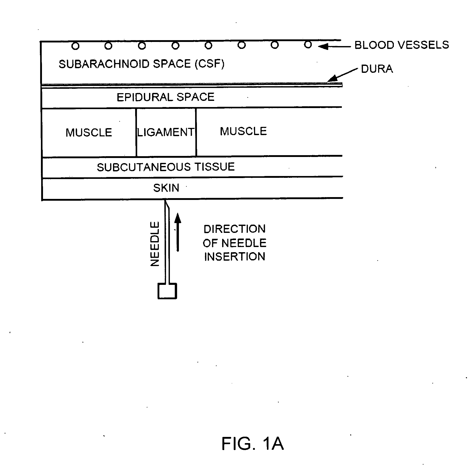
[0036]General aspects of a spinal canal access procedure and basic tissue anatomy are described with reference to FIGS. 1A through 1C. The spinal canal includes both the epidural space as well as the subarachnoid space containing CSF, with those two spaces separated by the dura. In an access procedure, a needle (or other probe) is inserted through the skin and subcutaneous tissue and into a tough ligament in the patient's back. In a lumbar puncture procedure, the needle advances through the ligament and into the epidural space, and further through the dura into the subarachnoid space for access to the CSF (e.g., FIGS. 1A and 1C). Precision and care is required in needle positioning, not on...


PUM


Abstract

Methods and devices for detecting positioning of a probe in a tissue of a patient. A method can include providing a detection device; advancing a device coupled probe through the tissue of the patient and toward the patient's spinal canal; detecting a change in pressure about the distal portion of the coupled probe during advancing, where the detected pressure change indicates probe positioning in the patient's spinal canal; outputting the detected pressure change or indication of probe positioning to a visual display.

Description

CROSS-REFERENCE TO RELATED APPLICATION[0001]The present invention claims the benefit of priority under 35 U.S.C. §119(e) of U.S. Provisional Application No. 61 / 235,004, filed Aug. 19, 2009 (Attorney Docket No. 028267-000100US) and U.S. Provisional Application No. 61 / 300,794, filed Feb. 2, 2010 (Attorney Docket No. 028267-000200US), the entire contents of which are herein incorporated by reference.[0002]The present application is related to U.S. application Ser. No. xx / xxx,xxx, filed Aug. 19, 2010 (Attorney Docket No. 028267-000300US), and U.S. application Ser. No. xx / xxx,xxx, filed Aug. 19, 2010 (Attorney Docket No. 028267-000400US), both of which are being filed concurrently herewith, the full disclosures of which are herein incorporated by reference.BACKGROUND OF THE INVENTION[0003]The present disclosure relates generally to systems, methods, and devices for facilitating access to a target anatomical site. More specifically, the present invention provides methods and structures fo...

Claims


Application Information

Patent Timeline
Patent Type & Authority Applications(United States)
IPC IPC(8): A61B5/03A61B17/34A61B5/06A61B5/00
CPCA61B17/3401A61B5/036A61B5/4821A61B5/065A61B5/032A61B2019/465A61M25/06A61M25/0693A61B2090/065A61B2090/034
Inventor HULVERSHORN, JUSTINSCHMIDT, KARLSWARTZ, DOUGLAS
Owner MEDLINE IND LP
Learn More