Method and device for imaging soft body tissue using x-ray projection and optical tomography

Active Publication Date: 2014-09-25
KONINKLIJKE PHILIPS ELECTRONICS NV
View PDF4 Cites 10 Cited by
  • Summary
  • Abstract
  • Description
  • Claims
  • Application Information

AI Technical Summary

Benefits of technology

The patent describes a method for improving the quality and resolution of optical tomography images by using X-ray projection image data to estimate the optical properties of soft body tissue and then using these estimates to improve the reconstruction of the optical tomography image. This method may be particularly useful for imaging female breasts where mammograms can be uncomfortable. The acquisition of third image data at a different compression state can further improve the quality of the second image. The proposed method allows for the combination of the advantages of different imaging techniques and improves the accuracy and reliability of optical tomography imaging.

Problems solved by technology

However, the proposed approach still suffers from shortcomings concerning e.g. image quality and / or patient comfort.

Method used


Image

  • Method and device for imaging soft body tissue using x-ray projection and optical tomography
  • Method and device for imaging soft body tissue using x-ray projection and optical tomography

Examples


Embodiment Construction

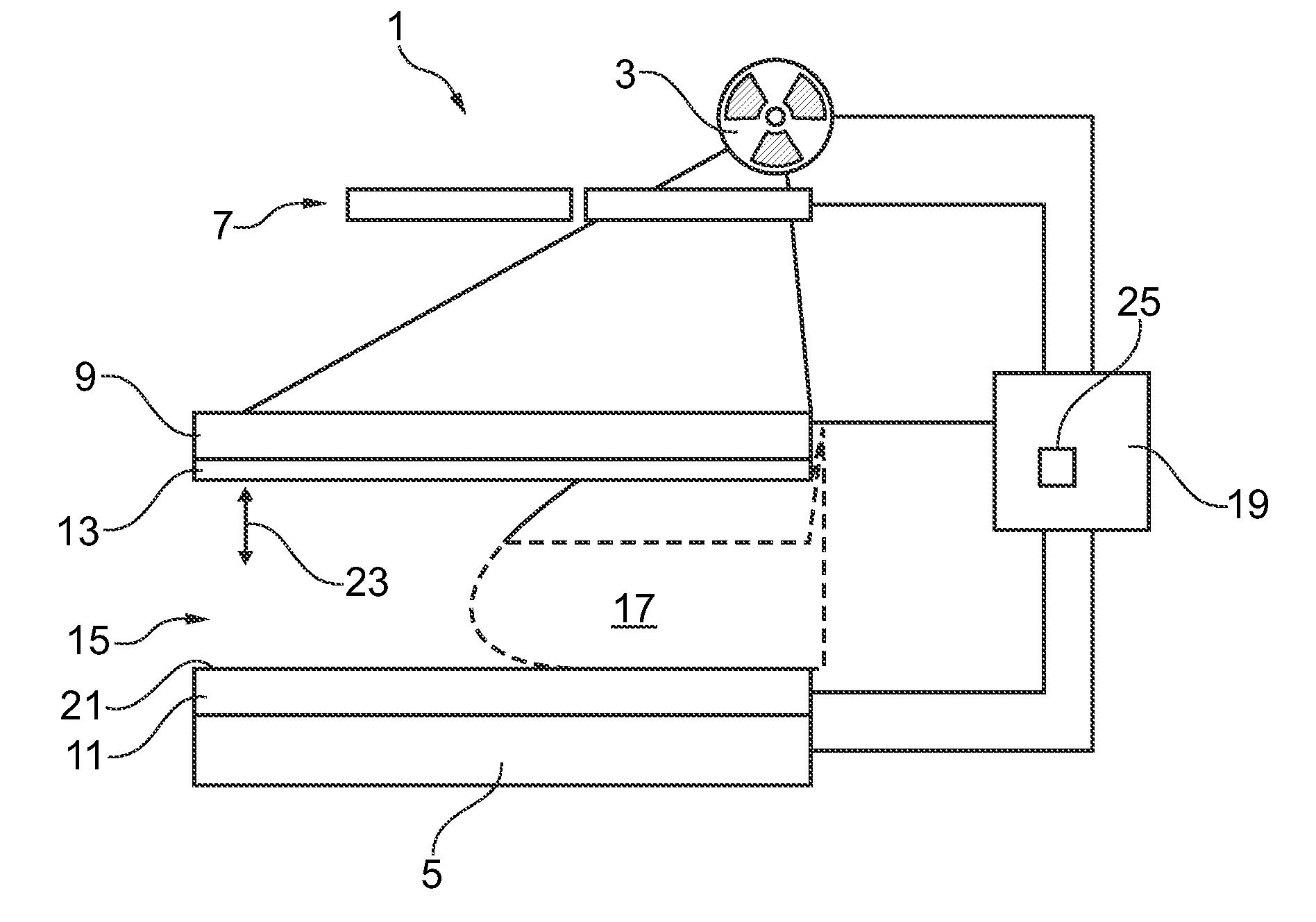
[0036]FIG. 1 shows an imaging device 1 which may be used for performing an imaging method according to embodiments of the present invention as described in further detail below. The imaging device 1 is adapted for imaging a female breast 17 at different compression states and with different imaging modalities.

[0037]The imaging device 1 comprises a soft body tissue compressor 15 having a compression paddle 13 which may be moved in a vertical direction 23 as indicated in FIG. 1. Using the movable compression paddle 13, a female breast 17 interposed between the compression paddle 13 and a housing 21 may be compressed to different compression states as schematically indicated in the figure by dotted and solid lines, respectively.

[0038]An X-ray source 3 and an X-ray detector 5 are arranged at opposite sides of the compressor 15. The X-ray source 3 may emit X-rays towards a region of interest within the breast 17 such that these X-rays are transmitted through the region of interest and ar...


PUM


Abstract

A method and device (1) for imaging soft body tissue such as a female breast is proposed. X-ray projection techniques and optical tomography techniques are combined. First image data for a first image of a breast (17) may be acquired by X-ray projection using an X-ray source (3) and an X-ray detector (5). Second image data for a second image may be acquired using optical tomography equipment comprising a light source (9) and a light detector (11). From the first image data, estimated bulk optical properties of the breast (17) are be derived. Based on such estimated bulk optical properties, an optical tomography image is reconstructed from the second image data with high image quality. Performing mammography acquisition at different compression states of the breast (17) may improve patient comfort. Mammograms may be acquired at two different compression states wherein a first compression state is adapted to provide high image resolution. At a second compression state, another mammogram may be acquired together with an optical tomography image. The two mammograms may be used for image registration thereby possibly providing information for a deformation prior. Additional information on tissue composition within the breast may be received by acquiring the first and second mammogram at different X-ray settings.

Description

FIELD OF THE INVENTION[0001]The present invention relates to a method and a device for imaging soft body tissue such as a female breast. Furthermore, the invention relates to a computer program product for performing such method and a computer-readable medium having stored thereon such computer program product.BACKGROUND OF THE INVENTION[0002]In order to be able to examine soft body tissue such as a female breast and possibly detect tumourous tissue therein, various imaging methods have been developed.[0003]For example, mammography imaging uses X-rays which are projected through the breast and are detected after transmission such that, from the detected X-ray intensity distribution, information may be derived with respect to geometry and X-ray absorption of structures within the breast. The images acquired by a mammography X-ray projection are typically two-dimensional (2D).[0004]As an alternative imaging technique, optical tomography has been developed. Optical tomography is a type...

Claims


Application Information

Patent Timeline
IPC IPC(8): A61B6/00A61B5/00
CPCA61B5/0035A61B6/4417A61B6/0414A61B6/463A61B6/482A61B6/502A61B6/5217A61B6/5247A61B6/54A61B5/0073A61B5/0091A61B5/7425G16H50/30
Inventor GOOSSEN, ANDREHEESE, HARALD SEPPKOEHLER, THOMAS
Owner KONINKLIJKE PHILIPS ELECTRONICS NV
Why Eureka
Try Eureka