Methods and compositions for identifying minimal residual disease in acute lymphoblastic leukemia
a lymphoblastic leukemia and minimal residual disease technology, applied in the field of minimal residual disease detection in patients with acute lymphoblastic leukemia, can solve the problems of undiagnosed relapse, limited application to specialized centers, and inability to detect the presence of leukemic cells, etc., and achieve the effect of diagnosing minimal residual diseas
- Summary
- Abstract
- Description
- Claims
- Application Information
AI Technical Summary
Benefits of technology
Problems solved by technology
Method used
Image
Examples
example 1
Identification of Genes Differentially Expressed in Leukemic and Normal Immature B Cells
[0196]Gene Expression Arrays Studies
[0197]A first-generation gene array study was previously performed (Chen et al. (2001) Blood. 97:2115-2120) and as previously described (Ross et al. (2003) Blood. 102:2951-2959). In Chen et al., diagnostic ALL samples from 4 patients were compared to CD19+ CD10+ bone marrow immature B cells taken from 2 healthy donors. The array contained probes for approximately 4000 genes.
[0198]In an embodiment of the invention, a gene expression array probing over 23,000 genes was used to screen a larger set of samples, including 270 cases of B-lineage ALL. The B-lineage ALLs encompassed the spectrum of genetic abnormalities occurring in ALL and were included to specifically identify other prognostic markers. Briefly, total RNA was isolated from freshly thawed B-lineage ALL cells and flow sorted normal CD19+CD10+ cells using the TRIZOL® reagent (Invitrogen, Carlsbad, Calif.)...
example 2
Validation of Gene Expression Array Results by Flow Cytometry
[0202]Among the genes differentially expressed by gene array analysis, there were some already widely used for MRD studies by flow cytometry, i.e., CD58, CD38, CD13, and CD34, suggesting the possibility that other useful markers could be present among the remaining genes (Basso et al. (2009) J Clin Oncol. 27:5168-5174; Lucio et al. (2001) Leukemia. 15:1185-1192; Chen et al. (2001) Blood. 97:2115-2120; Coustan-Smith et al. (2002) Blood. 100:52-58).
[0203]To prioritize genes for validation by flow cytometry, an initial inclusion criteria was applied: a) differential expression in at least 25% of cases of ALL, or 40% of cases of a genetic subtype of ALL; b) over-expression in leukemic cells by at least 3-fold of the maximum value in normal cells, or under-expression by 3-fold of the minimum value in normal cells; and c) commercial availability of specific antibodies conjugated to fluorochromes suitable for flow cytometry. Guid...
example 3
Validation of the New Markers for MRD Detection
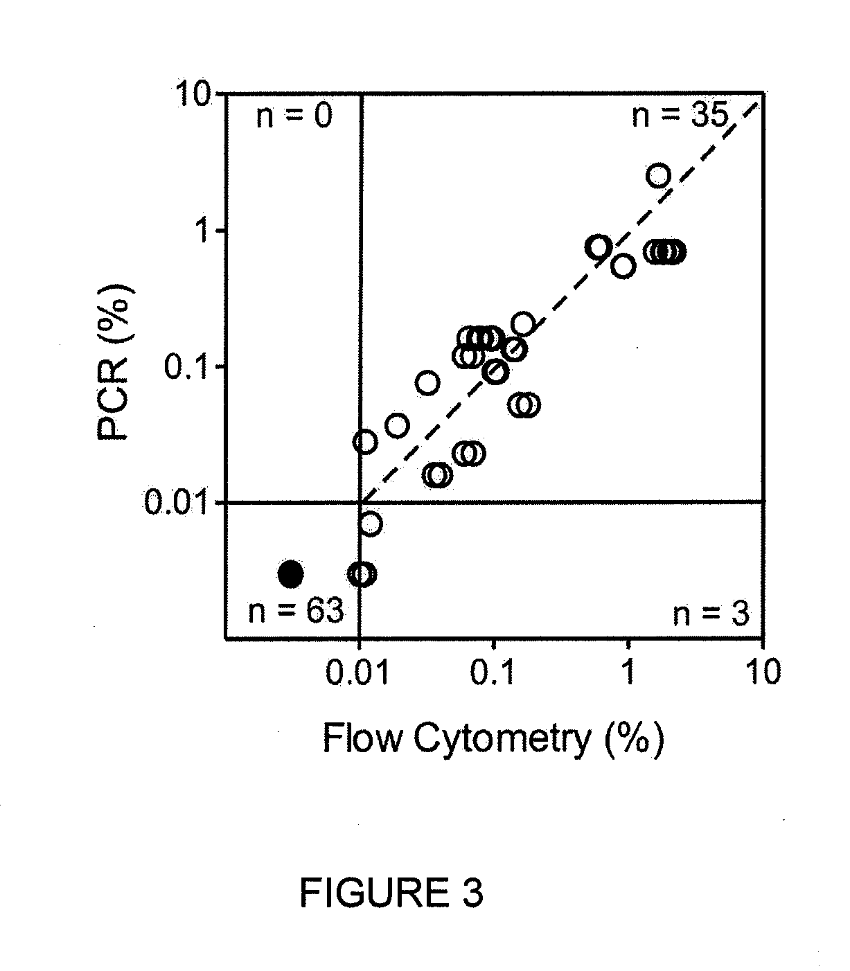
[0209]To determine the reliability of the new markers to identify leukemic cells in clinical samples, 128 bone marrow samples were collected during treatment (46 during or at the end of remission induction therapy and 82 during post-remission therapy) from 51 patients with B-lineage ALL in whom expression of the markers on the leukemic cells had been measured at diagnosis. The markers included in these studies were the top 16 differentially expressed markers listed in TABLE 7, for a total of 258 tests.
[0210]Newly identified markers (TABLE 3, 4) were compared to standard marker combinations presently used in for MRD detection (TABLE 5). Using a threshold of 0.01% ALL cells to define MRD positivity, no discordant results were observed except for one comparison in which MRD was negative with the standard markers and 0.012% with the new markers. Overall, there was an excellent correlation in MRD estimates between new and standard markers (r...
PUM
| Property | Measurement | Unit |
|---|---|---|
| Level | aaaaa | aaaaa |
Abstract
Description
Claims
Application Information
 - R&D
- Intellectual Property
- Life Sciences
- Materials
- Tech Scout
- Unparalleled Data Quality
- Higher Quality Content
- 60% Fewer Hallucinations
Browse by: Latest US Patents, China's latest patents, Technical Efficacy Thesaurus, Application Domain, Technology Topic, Popular Technical Reports.
© 2025 PatSnap. All rights reserved.Legal|Privacy policy|Modern Slavery Act Transparency Statement|Sitemap|About US| Contact US: help@patsnap.com


