
Noninvasive detection of fetal genetic abnormality

a fetal genetic and noninvasive technology, applied in the field of noninvasive methods for the detection of fetal genetic abnormalities, can solve the problems of invasive prenatal diagnostic methods such as chorionic villus sampling and amniocentesis, carry potential risks for both fetuses and mothers, and noninvasive screening of fetal aneuploidy using maternal serum markers and ultrasound, and have limited sensitivity and specificity. , the diagnostic and clinical application of circulating

Active Publication Date: 2014-04-10
BGI GENOMICS CO LTD
View PDF2 Cites 12 Cited by
  • Summary
  • Abstract
  • Description
  • Claims
  • Application Information

AI Technical Summary

Benefits of technology

The patent text describes methods for non-invasive detection of fetal genetic abnormalities by analyzing nucleotide sequences from a maternal sample. The methods involve assigning polynucleotide fragments to chromosomes and calculating coverage depth and GC content of chromosomes. The relationship between coverage depth and GC content is then used to determine if there is a difference between the two, indicating fetal genetic abnormality. The methods may be used with samples from pregnant female or male subjects and can be performed using parallel genomic sequencing or other genomic sequencing techniques. The patent text also provides a formula for estimating fetal fraction and a formula for determining fetal gender.

Problems solved by technology

Conventional prenatal diagnostic methods with invasive procedures, such as chorionic villus sampling and amniocentesis, carry potential risks for both fetuses and mothers.
Noninvasive screening of fetal aneuploidy using maternal serum markers and ultrasound are available but have limited sensitivity and specificity (Kagan, et al., Human Reproduction (2008) 23:1968-1975; Malone, et al., N Engl J Med (2005) 353:2001-2011).
However, the diagnostic and clinical applications of circulating fetal DNA is limited to genes that are present in the fetus but not in the mother (Pertl and Bianchi, Obstetrics and Gynecology (2001) 98:483-490).
However, as some studies show, GC bias introduced by amplification and sequencing placed a practical limit on the sensitivity of aneuploidy detection.
However, the method has difficulty in dealing with sex chromosome disorders especially chromosome Y relevant disorders for the reason that the process may cause slight distortion of data which will interfere with the precision of detection.

Method used


Image

  • Noninvasive detection of fetal genetic abnormality
  • Noninvasive detection of fetal genetic abnormality
  • Noninvasive detection of fetal genetic abnormality

Examples


example 1

Analysis of Factors that Affect Sensitivity of Detection: GC-Bias and Gender

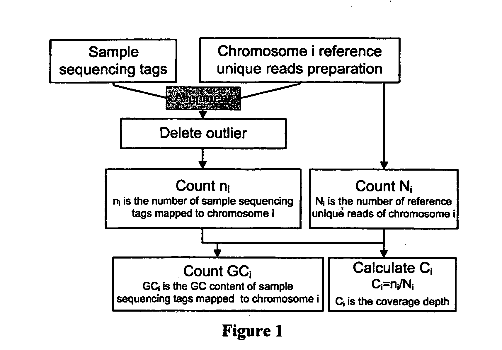
[0084]A schematic procedural framework for calculating coverage depth and GC content is illustrated in FIG. 1. We used software to produce the reference unique reads by incising the hg18 reference sequences into 1-mer (1-mer here is a read being artificially decomposed from the human sequence reference with the same “1” length with sample sequencing reads) and collected those “unique” 1-mer as our reference unique reads. Secondly, we mapped our sequenced sample reads to the reference unique reads of each chromosome. Thirdly, we deleted the outlier by applying quintile outlier cutoff method to get a clear data set. Finally, we counted the coverage depth of each chromosome for every sample and the GC content of the sequenced unique reads mapped to each chromosome for every sample.

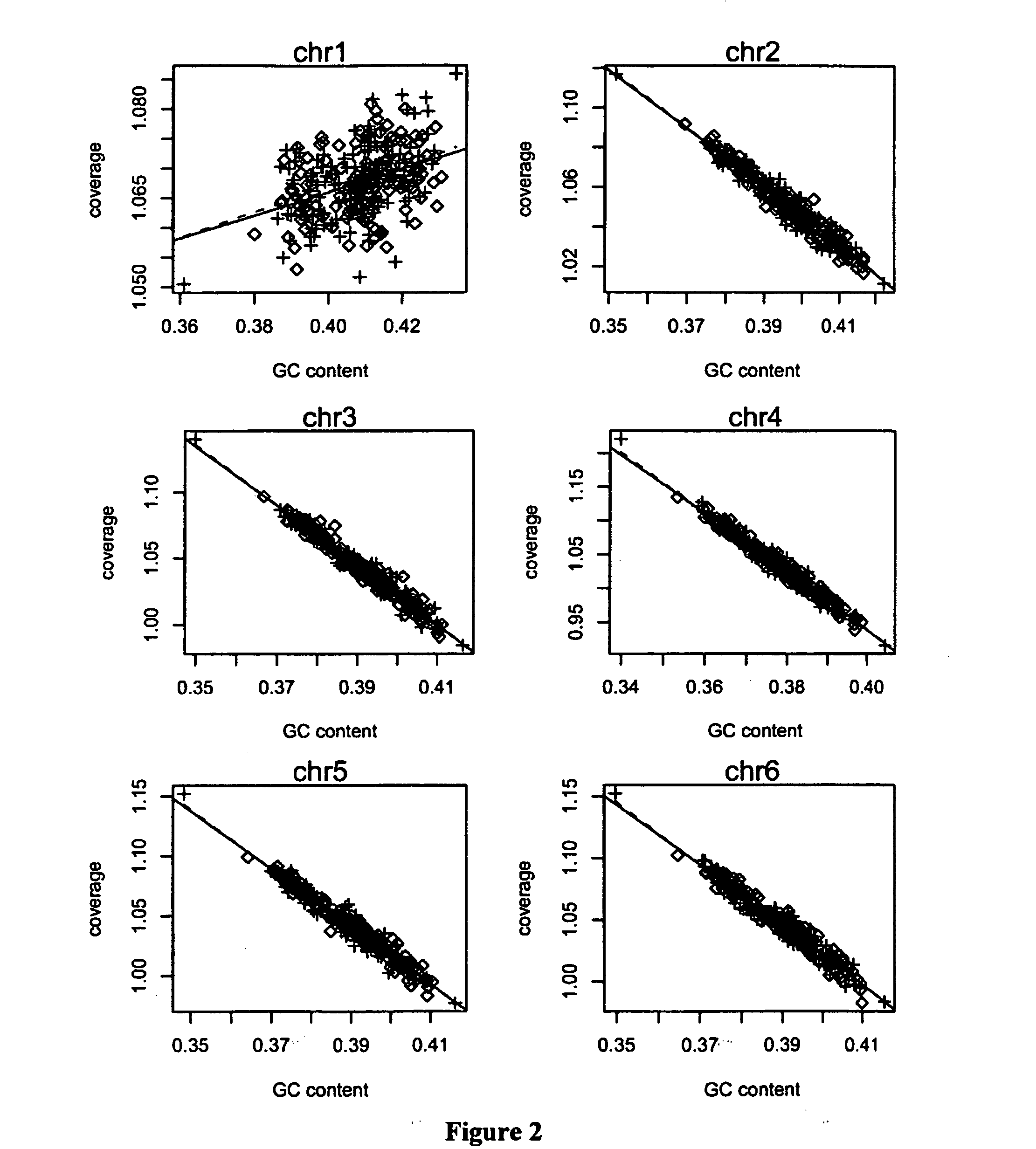
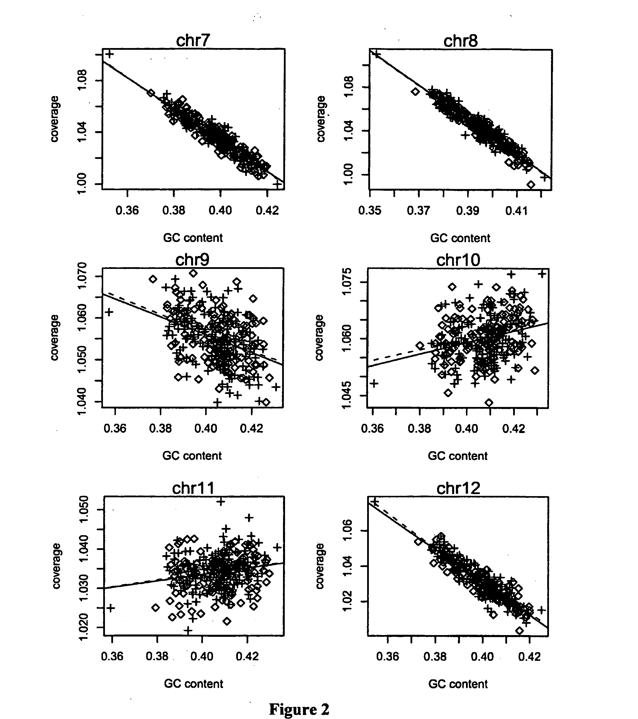
[0085]In order to investigate how GC content affects our data, we chose 300 euploid cases with karyotype result and scattered their ...

example 2

Statistical Model

[0088]Using this phenomenon discussed above, we tried to use local polynomial to fit the relationship between coverage depth and the corresponding GC content. The coverage depth consists of a function of GC and a residual of normal distribution as following:

cri,j=ƒ(GCi,j)+εi,j, j=1,2, . . . , 22,X,Y  (4)

wherein ƒ(GCi,j) represents the function for the relationship between coverage depth and the corresponding GC content of sample i, chromosome j, εi,j represents the residual of sample i, chromosome j.

[0089]There is non-strong linear relationship between the coverage depth and the corresponding GC content so we applied loess algorithm to fit the coverage depth with the corresponding GC content, from which we calculated a value important to our model, that is, the fitted coverage depth:

c{circumflex over (r)}i,j=ƒ(GCi,j), j=1,2, . . . , 22,X,Y  (5)

With the fitted coverage depth, the standard variance and the student t were calculated according to the flowing Formula 6 a...

example 3

Fetal Fraction Estimation

[0090]For the reason that fetal fraction is very important for our detection so we estimated the fetal fraction before the testing procedure. As we had mentioned before, we had sequenced 19 male adults, when compared their coverage depth with that of cases carrying female fetus, we found that male's coverage depth of chromosome X is almost ½ times of female's, and male's coverage depth of chromosome Y is almost 0.5 larger than female's. Then we can estimate the fetal fraction depending on the coverage depth of chromosome X and Y as Formula 8, Formula 9 and Formula 10, considering GC-correlation as well:

fyi=(cri,Y-c^ri,Yf) / (c^ri,Ym-c^ri,Yf)(8)fxi=(cri,X-c^ri,Xf) / (c^ri,Xm-c^ri,Xf)(9)fxyi=argminɛ∈(0,1)((c^ri,Xf·(1-ɛ)+c^ri,Xm·ɛ-cri,X)2(σ^X,f·(1-ɛ))2+(σ^X,m·ɛ)2+(c^ri,Yf·(1-ɛ)+c^ri,Ym·ɛ-cri,X)2(σ^Y,f·(1-ɛ))2+(σ^Y,m·ɛ)2)(10)

wherein ĉri,Xf=ƒ(GCi,Xf) is the fitted coverage depth by the regression correlation of the chromosome X coverage depth and corresponding GC con...


PUM

PropertyMeasurementUnit
coverage depthaaaaaaaaaa
depthaaaaaaaaaa
conditional probability densityaaaaaaaaaa

Abstract

The current invention is directed to methods for noninvasive detection of fetal genetic abnormalities by large-scale sequencing of nucleotides from maternal biological sample. Further provided are methods to remove GC bias from the sequencing results according to the difference in GC content of a chromosome. The current invention not only makes the detection much more accurate but also represents a comprehensive method for fetal aneuploidy detection including sex chromosome disorders such as XO, XXX, XXY, and XYY, etc.

Description

TECHNICAL FIELD[0001]The invention relates to noninvasive methods for the detection of fetal genetic abnormality by DNA sequencing of samples from pregnant women. More particularly, this invention relates to data analysis to remove GC bias introduced by amplification and sequencing of DNA samples. This invention further relates to statistical analysis to detect fetal genetic abnormalities, such as chromosomal abnormalities including aneuploidy.BACKGROUND ART[0002]Conventional prenatal diagnostic methods with invasive procedures, such as chorionic villus sampling and amniocentesis, carry potential risks for both fetuses and mothers. Noninvasive screening of fetal aneuploidy using maternal serum markers and ultrasound are available but have limited sensitivity and specificity (Kagan, et al., Human Reproduction (2008) 23:1968-1975; Malone, et al., N Engl J Med (2005) 353:2001-2011).[0003]Recent studies have demonstrated noninvasive detection of fetal aneuploidy by massively parallel se...

Claims


Application Information

Patent Timeline
Patent Type & Authority Applications(United States)
IPC IPC(8): G06F19/18G16B20/10G16B20/20G16B30/10
CPCG06F19/18C12Q1/6883C12Q2600/156G16B20/00G16B30/00G16B20/10G16B20/20G16B30/10C12Q1/6844
Inventor JIANG, FUMANCHEN, HUIFEICHAI, XIANGHUAYUAN, YUYINGZHANG, XIUQINGCHEN, FANG
Owner BGI GENOMICS CO LTD
Learn More