
Minimally Invasive Endoscopic/Laparoscopic Highly Absorbent Surgical Devices, Methods and System

Inactive Publication Date: 2014-03-27
BENGTSON BRADLEY P
View PDF10 Cites 34 Cited by
  • Summary
  • Abstract
  • Description
  • Claims
  • Application Information

AI Technical Summary

Benefits of technology

The present invention is a sponge device that can be used during endoscopic and laparoscopic procedures. The device can be introduced through a disposable sleeve and is designed to absorb blood and body fluids. It can also be used as a drain and help with coagulation. The sponge device can be easily placed and removed and can help initiate clotting. Overall, the invention provides a useful tool for surgeons to improve the procedures' efficiency and effectiveness.

Problems solved by technology

Currently there is no sponge available for intraoperative minimally invasive or laparoscopic assisted procedures.
As with other surgical procedures, the removal of bodily fluids, such as irrigation fluid introduced by the surgeon, blood, exudates and all blood products during the procedure is a concern for minimally invasive surgery as it is for open surgery with any of these blood or body fluids decreasing visualization for the surgeon, which interferes with the operation.
However, such materials are not necessarily conducive for use in minimally invasive surgery, as the requirement of inserting the material through the cannula is not the easiest task to perform.
In addition the current sponges are not very absorbent, can fragment and also carry a great deal of lint and surgical debris into the body.
Raytec® or other sponge products, or Kittner dissectors may be placed through a cannula, but are not typically used or have significant limitations because of multiple issues described above.
These products fragment, leave debris, are difficult to remove, and do not transmit any fluid or allow suction of blood or fluid through them.
These sponges are incapable of fluid absorption and must be replaced frequently, and again may fragment.
In addition they tend to adhere or stick to open surgical wounds or dissection sites and then may cause additional bleeding when removed verses our new device which is absorbent but also non-adherent.
These other devices are essentially a rolled up cotton sponges, and although they may be helpful to do blunt dissection, they are inadequate to do all that is required for fluid transduction and removal, and are more commonly used to dissect tissues verses a true sponge utilized in an open procedure.

Method used


Image

  • Minimally Invasive Endoscopic/Laparoscopic Highly Absorbent Surgical Devices, Methods and System
  • Minimally Invasive Endoscopic/Laparoscopic Highly Absorbent Surgical Devices, Methods and System
  • Minimally Invasive Endoscopic/Laparoscopic Highly Absorbent Surgical Devices, Methods and System

Examples


embodiment 20

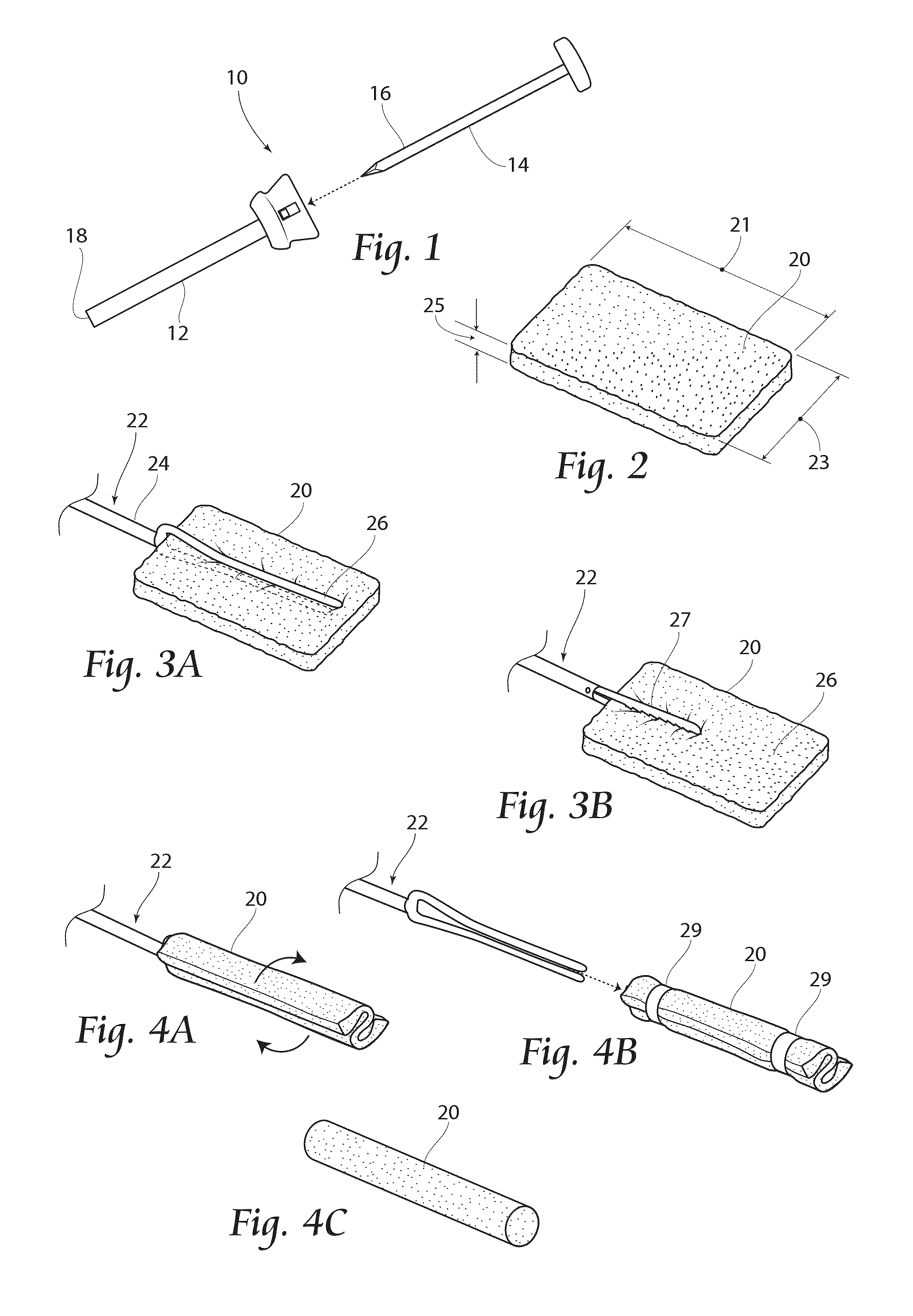
[0059]FIG. 4C provides an alternate embodiment 20″ of a sponge cartridge unit, including a unitary sponge device 20 of generally cylindrical shape. Any of the cartridge units 20′,20″ may be used alone or in conjunction with an introducer device, such as that shown in FIGS. 11 and 12, and described below.

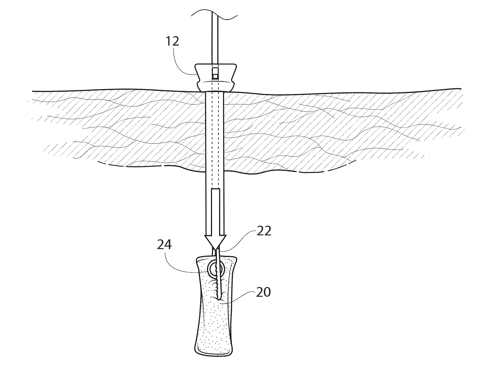
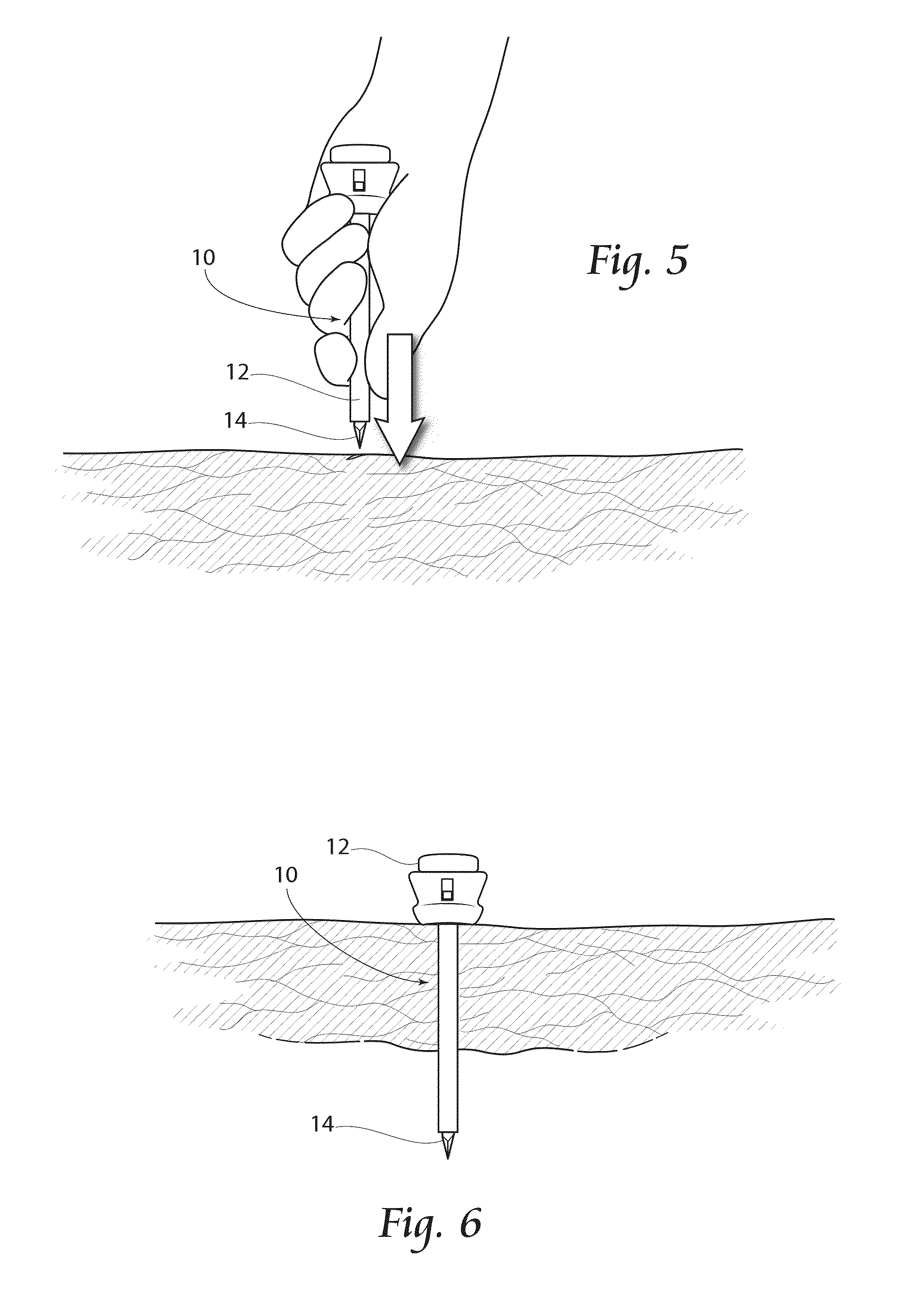
[0060]Referring now to FIGS. 5 and 6, the assembly 10 is shown being inserted into tissue and into a body cavity. The sharpened end of the trocar 14 is pushed downwardly into the tissue and eventually extending into the body cavity, as shown in FIG. 6. Once inserted into the body cavity to a desired depth, the trocar 14 is removed from the assembly 10, with the cannula 12 remaining in place to allow a pathway into the body cavity.

[0061]As shown in FIG. 7, the absorbent device 20 is being inserted into the cannula 12 by way of the insertion device 22, as described above. The absorbent device 20 is in the compact, cylindrical position, which allows it to pass through the cannula 12, in...

first embodiment

[0079]FIG. 34 depicts a sponge kit 300 according to the present invention. The kit 300 preferably includes a container 302, such as a draw-formed thermoplastic tray, which may form one or more compartments 304. The compartments 304 are preferably sealable by, e.g., plastic film (not shown) which may be adhered to the tray 302. This first kit 300 includes a cannula 12, a trocar 14, a sponge retraction tool 22, one or more sponge devices 20, and instructions 306 for using the sponge 20 in conjunction with the provided system components. The instructions for use 306 generally would set forth the method steps described herein. Each provided component may be placed in its own compartment 304, or a plurality of components may share a compartment 304. Additionally, a kit 300′ may include a sponge introducer 100, as shown in FIG. 35. Additionally or alternatively, the cannula 12 and trocar 14 may be eliminated from a kit 400, and / or a plurality of sizes of sponges 20 may be provided, as sho...


PUM


Abstract

Devices, systems and methods related to fluid control during laparoscopic surgical methods are disclosed. A device includes a highly absorbent surgical sponge device comprising highly absorbent material capable of being inserted through a port or cannula, which has been inserted into a body cavity. The device may include a fluid conduit coupled to the absorbent pad to remove fluids from or introduce fluids into the body cavity through the device during the course of surgery.

Description

RELATED APPLICATIONS[0001]This application claims the benefit of co-pending U.S. Provisional Patent Application Ser. No. 61 / 706,393, filed Sep. 27, 2012, and entitled “Endoscopic / Laparoscopic Absorbent Devices, Methods and System,” which is incorporated herein by reference.BACKGROUND OF THE INVENTION[0002]Currently, up to 30% of general surgical, and many subsurgical, procedures employ an endoscope. All current open procedures utilize sponges of some sort to facilitate the surgical procedure. Currently there is no sponge available for intraoperative minimally invasive or laparoscopic assisted procedures. The present invention relates to the design and use of a family of novel, intraoperative, highly absorbent surgical sponge devices, and, more particularly, to the design and use of surgical sponge devices utilized in minimally invasive or laparoscopic surgical procedures, including the specific unique new methods and accompanying systems.[0003]Minimally invasive surgery, including l...

Claims


Application Information

Patent Timeline
IPC IPC(8): A61F13/36
CPCA61F13/36
Inventor BENGTSON, BRADLEY, P.
Owner BENGTSON BRADLEY P
Learn More