
System and method for detecting critical structures using ultrasound

a technology of critical structures and ultrasound, applied in the field of open or endoscopic surgical instruments and methods, can solve the problems of poor visibility, inability to detect critical structures, and most difficult tasks, and achieve the effects of low cost, effective integration, and reduced volume of information provided to surgeons

Active Publication Date: 2014-01-16
TYCO HEALTHCARE GRP LP
View PDF2 Cites 64 Cited by
  • Summary
  • Abstract
  • Description
  • Claims
  • Application Information

AI Technical Summary

Benefits of technology

This patent describes a way to create a laparoscopic device that has a single ultrasound transducer. This transducer is easy to integrate and helps reduce the amount of information provided to the surgeon. It only provides a single perspective on tissue data, which is useful in improving the precision of surgical procedures. This technology is cost-effective and can help make minimally invasive surgeries even more minimally invasive.

Problems solved by technology

Smaller cannulae are usually preferred, which, as can be appreciated, ultimately presents a design challenge to instrument manufacturers who must find ways to make surgical instruments that fit through the cannulae.
This task is most difficult when the target anatomy (or that to be avoided) is obscured by overlying soft tissue.
One issue during laparoscopic procedures in particular, is inadvertently injuring nearby critical anatomical structures due to quick or abrupt movement of instruments within the surgical site, poor visibility, lack of tactile response, confusion of the anatomy from patient to patient, or inadequate control of the instrumentation being utilized to perform the procedure.
Unfortunately, the ureter is at times obscured by connective tissue, and difficult to identify.
The placement in such delicate structures is quite difficult and time consuming.
The use of an additional surgeon, one who likely is not otherwise involved in the underlying procedure being performed, significantly increases the cost of the procedure being performed.
Further the placement is time consuming and thus increases the overall operating time for the patient.
Again this increases the costs of the procedure, as well as increasing the time the patient must be sedated.
With respect specifically to the detection of the ureter, placement of the stent or catheter can be quite problematic.
However, these visualization techniques are often not conveniently useable to identify structures while the surgical procedures described above are on-going.
Further, the radiation levels imparted on the patient as well as clinicians is considered inherently safe which is not necessarily true for CT.
There are however, disadvantages to current to ultrasound techniques, particularly the (laparoscopic approaches) which have limited its adoption in minimally invasive surgery.
While this may be useful in the diagnostic phase of care, this is likely more information than is necessary in the real-time treatment venue of an operating room where the questions facing the surgeon are not “What's wrong?” but simply, “Where is it?” Still further in many surgical suites, the required ultrasound cart and probes are simply not available as the need for sub-surface imaging does not exist in every surgical case.
One drawback of current imaging systems is the size and cost of linear imaging arrays comprised of many small piezo electric elements.
While they provide detailed images, these probes are costly to manufacture and bulky relative to the size of laparoscopic surgical devices.
Further, according to another aspect the information presented to the surgeon is limited to just the most relevant information regarding subsurface structures rather than a full diagnostic image.

Method used


Image

  • System and method for detecting critical structures using ultrasound
  • System and method for detecting critical structures using ultrasound
  • System and method for detecting critical structures using ultrasound

Examples


Embodiment Construction

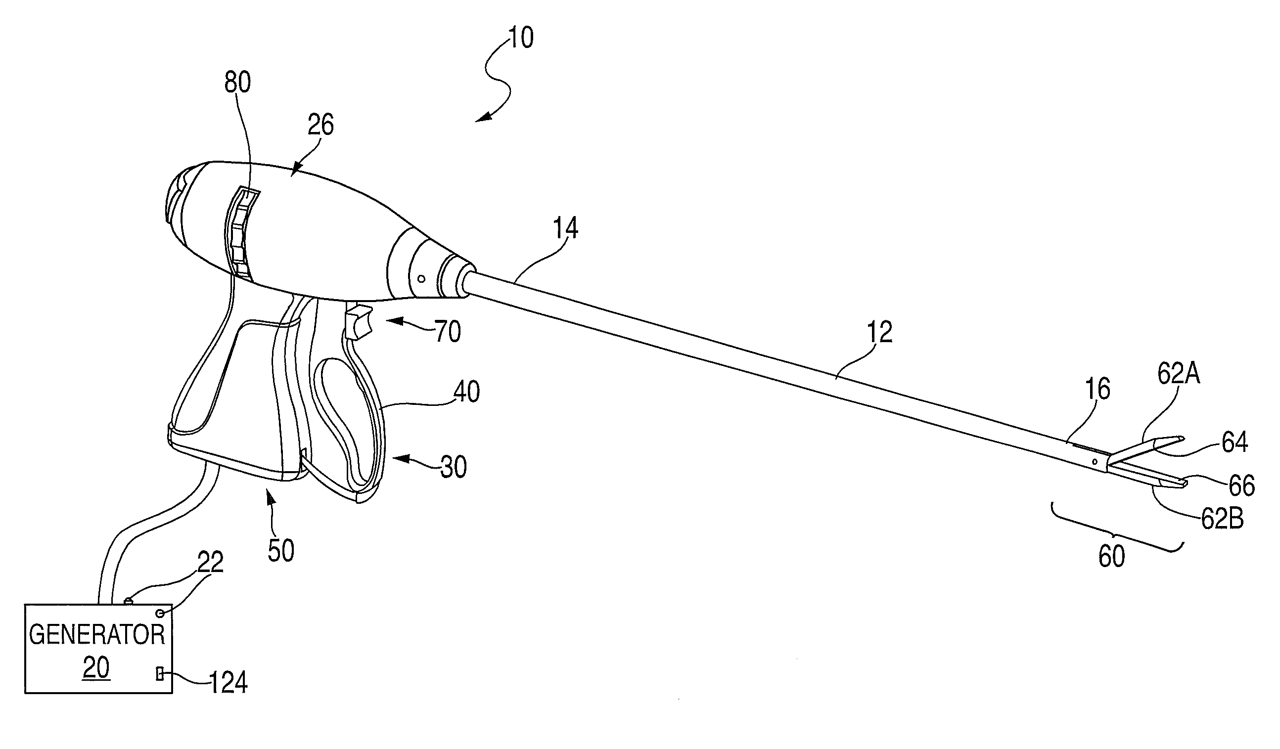
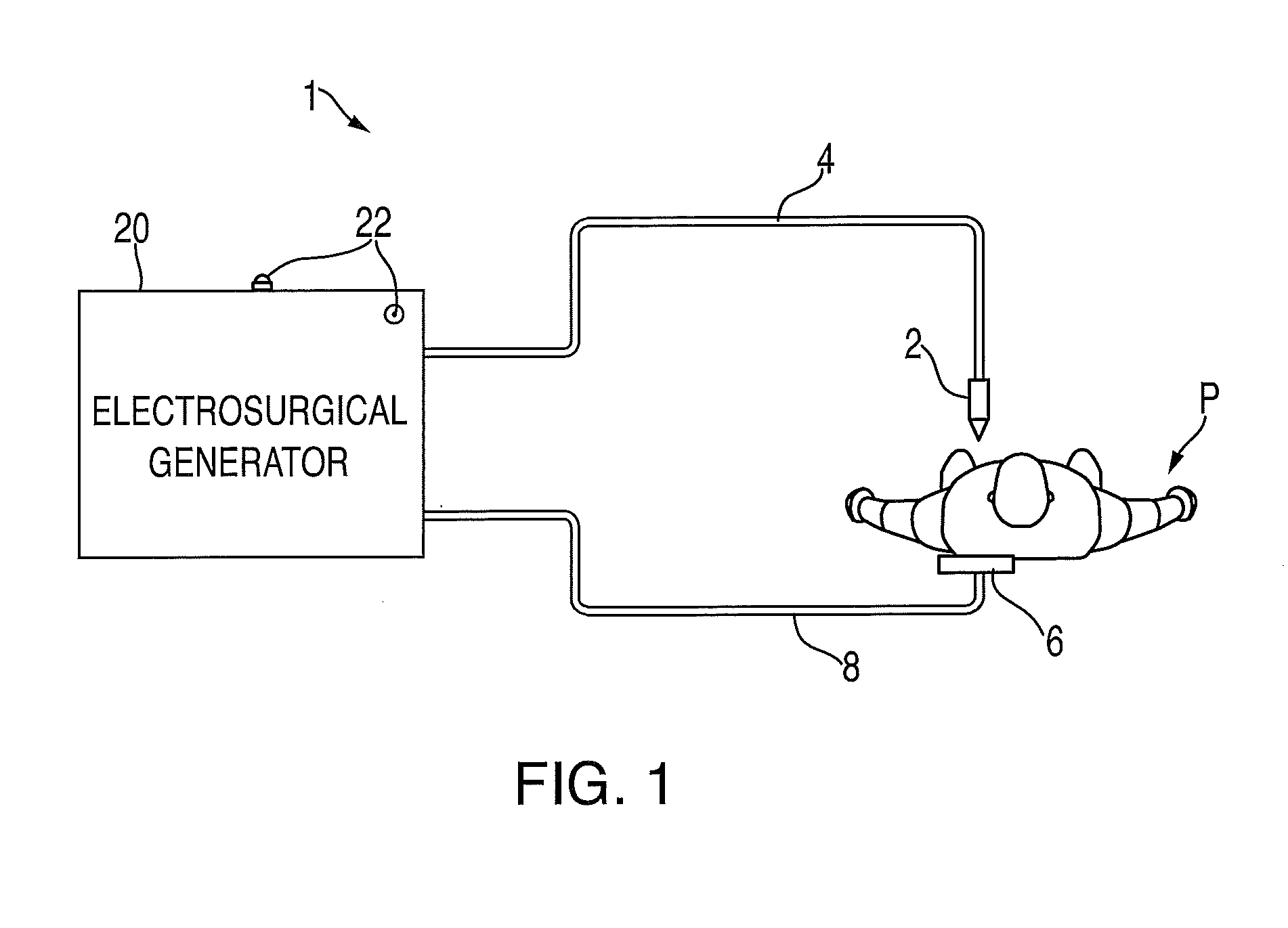
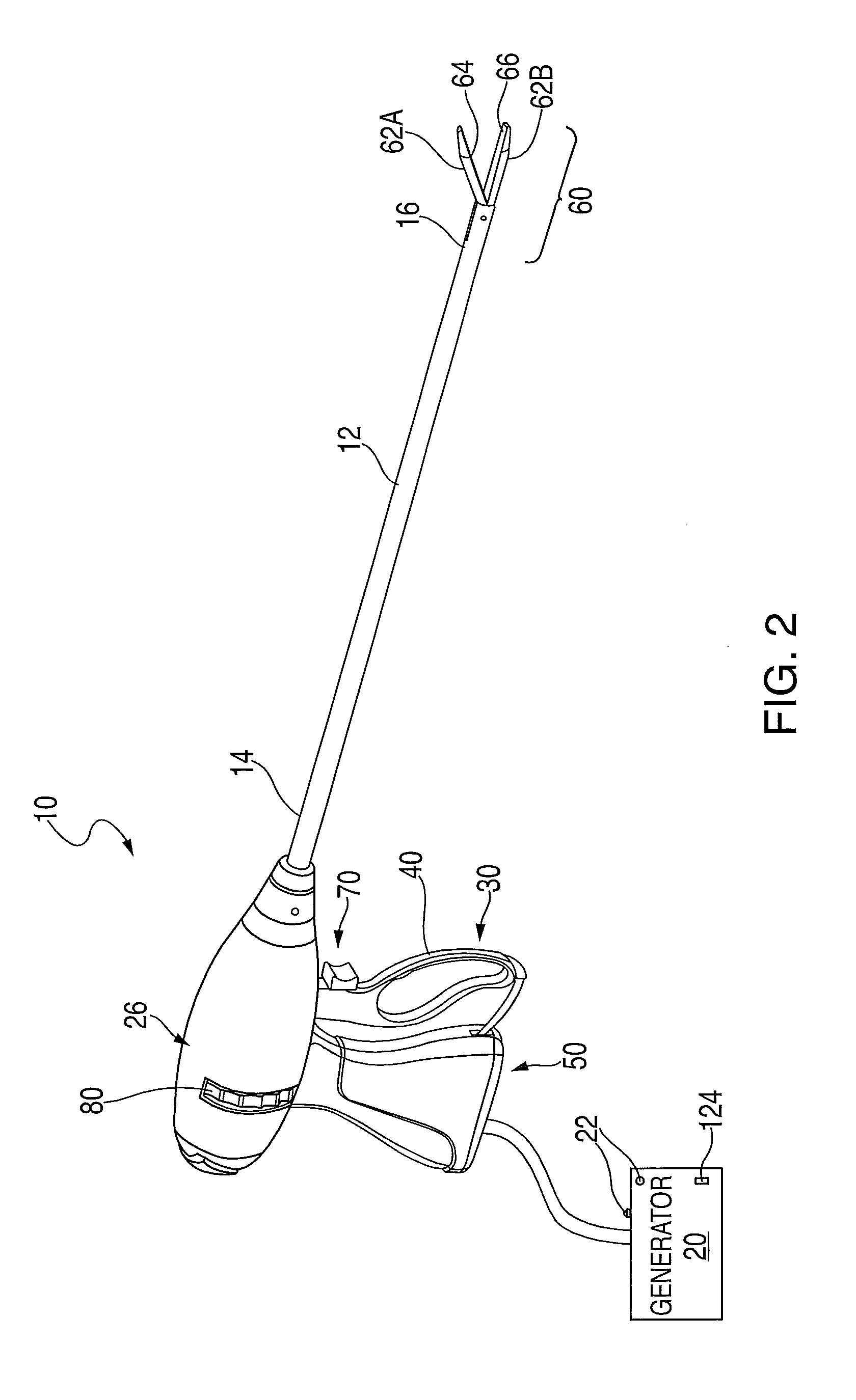
[0060]Particular embodiments of the present disclosure are described with reference to the accompanying drawings. In the following description, well-known functions or constructions are not described in detail to avoid obscuring the present disclosure in unnecessary detail. As used herein, the term distal and proximal are with respect to the medical professional utilizing the device or component, with proximal being nearer to the medical professional either when in use or during insertion to the patient. Thus, for example, the distal end of a surgical instrument is that portion nearer the end which will be inserted into a patient and the proximal end is that portion nearer the end which will remain outside the patient, and be available for treatment application by the medical professional.

[0061]The present disclosure generally relates to a system for identifying anatomical structures, particularly a lumen such as, for example, a ureter, bile or gall duct, a blood vessel, lymph notes...


PUM


Abstract

A system and method of ultrasound imaging, the system including a laparoscopic surgical instrument including at least one ultrasound transducer, and a processor adapted to receive acoustic data from the ultrasound transducer and process the acoustic data from the ultrasound transducer to produce a graphical representation of the acoustic data, the graphical representation depicting echoic attributes of tissue substantially axially aligned with a transmission path of the at least one ultrasound transducer, and the distance of at least one tissue type having different echoic attributes from surrounding tissue form a distal tip of the laparoscopic surgical instrument.

Description

CROSS REFERENCE TO RELATED APPLICATION[0001]The present application claims the benefit of and priority to U.S. Provisional Application Ser. No. 61 / 670,713, filed on Jul. 12, 2012, the entire contents of which are incorporated herein by reference.TECHNICAL FIELD[0002]The present disclosure relates to open or endoscopic surgical instruments and methods for treating tissue. More particularly, the present disclosure relates to a system and method for medical imaging and detecting critical structures and their proximity to a surgical device.BACKGROUND OF RELATED ART[0003]A hemostat or forceps is a simple pliers-like tool that uses mechanical action between its jaws to constrict vessels and is commonly used in open surgical procedures to grasp, dissect and / or clamp tissue. Electrosurgical forceps utilize both mechanical clamping action and electrical energy to affect hemostasis by heating the tissue and blood vessels to coagulate, cauterize and / or seal tissue.[0004]Over the last several d...

Claims


Application Information

Patent Timeline
Patent Type & Authority Applications(United States)
IPC IPC(8): A61B8/00
CPCA61B8/4254A61B8/12A61B1/3132A61B8/085A61B8/0891A61B8/4245A61B8/445A61B8/461A61B8/486A61B8/488A61B8/5215A61B8/5223A61B18/1482
Inventor ZHENG, PENGVALENTINE, CHRISTOPHER A.RUSIN, CHRISTOPHER T.ROOKS, KATHY E.CORUM, BENJAMIN M.JANDRALL, SALLY D.JOHNSON, KRISTIN D.
Owner TYCO HEALTHCARE GRP LP
Learn More