
Interventional In-Situ Image-Guidance by Fusing Ultrasound and Video

a multi-modality fusion and ultrasound technology, applied in the field of imaging devices and augmentation devices, can solve the problems of insufficient navigation accuracy, inconvenient use, and inability to natively assist multi-modality fusion, and achieve the effect of improving alignment performance and improving alignment performance for users

Inactive Publication Date: 2013-08-22
CLEAR GUIDE MEDICAL
View PDF6 Cites 209 Cited by
  • Summary
  • Abstract
  • Description
  • Claims
  • Application Information

AI Technical Summary

Benefits of technology

This patent is about a way to make ultrasound images easier for people to see and use for medical purposes. It uses a technique called adaptive projection to overcome interference and make the image clearer. This can be useful for people who need to use ultrasound for diagnostic or interventional purposes. The patent also mentions that it can help people who are learning about ultrasound or who need to use it for treatment purposes. Overall, this technique improves the alignment performance of ultrasound images and makes them easier for people to use.

Problems solved by technology

Limitations of the current approach on both the research and commercial sides can be attributed to the available tracking technologies and to the feasibility of integrating these systems and using them in clinical environments.
For example, mechanical-based trackers are considered expensive and intrusive solutions, i.e. they require large space and limit user motion.
Acoustic tracking does not provide sufficient navigation accuracy, leaving optical and EM tracking as the most successful and commercially available tracking technologies.
However, both technologies require intrusive setups with a base camera (in case of optical tracking methods) or a reference EM transmitter (in case of EM methods).
Furthermore, none of these systems natively assist multi-modality fusion (registration e.g. between pre-operative CT / MRI plans and intra-operative ultrasound), and do not contribute to direct or augmented visualization either.

Method used


Image

  • Interventional In-Situ Image-Guidance by Fusing Ultrasound and Video
  • Interventional In-Situ Image-Guidance by Fusing Ultrasound and Video
  • Interventional In-Situ Image-Guidance by Fusing Ultrasound and Video

Examples


example 1

Ultrasound-Guided Liver Ablation Therapy

[0270]Recent evidence suggests thermal ablation in some cases can achieve results comparable to that of resection. Specifically, a recent randomized clinical trial comparing resection to RFA for small HCC found equivalent long-term outcomes with lower morbidity in the ablation arm [Chen-2006] Importantly, most studies suggest that efficacy of RFA is highly dependent on the experience and diligence of the treating physician, often associated with a steep learning curve [Poon-2004]. Moreover, the apparent efficacy of open operative RFA over a percutaneous approach reported by some studies suggest that difficulty with targeting and imaging may be contributing factors [Mulier-2005]. Studies of the failure patterns following RFA similarly suggest that limitations in real-time imaging, targeting, monitoring of ablative therapy are likely contributing to increased risk of local recurrence [Mulier-2005].

[0271]One of the most useful features of ablativ...

example 2

Monitoring Neo-Adjuvant Chemotherapy Using Advanced Ultrasound Imaging

[0294]Out of more than two hundred thousand women diagnosed with breast cancer every year, about 10% will present with locally advanced disease [Valero-1996]. Primary chemotherapy (a.k.a. Neo-adjuvant chemotherapy, NAC) is quickly replacing adjuvant (post-operative) chemotherapy as the standard in the management of these patients. In addition, NAC is often administered to women with operable stage II or III breast cancer [Kaufmann-2006]. The benefit of NAC is two fold. First, NAC has the ability to increase the rate of breast conserving therapy. Studies have shown that more than fifty percent of women, who would otherwise be candidates for mastectomy only, become eligible for breast conserving therapy because of NAC induced tumor shrinkage [Hortabagyi-1988, Bonadonna-1998]. Second, NAC allows in vivo chemo-sensitivity assessment. The ability to detect early drug resistance will prompt change from the ineffective t...

example 3

Ultrasound Imaging Guidance for Laparoscopic Partial Nephrectomy

[0313]Kidney cancer is the most lethal of all genitourinary tumors, resulting in greater than 13,000 deaths in 2008 out of 55,000 new cases diagnosed [61]. Further, the rate at which kidney cancer is diagnosed is increasing [1,2,62]. “Small” localized tumors currently represent approximately 66% of new diagnoses of renal cell carcinoma [63].

[0314]Surgery remains the current gold standard for treatment of localized kidney tumors, although alternative therapeutic approaches including active surveillance and emerging ablative technologies [5] exist. Five year cancer-specific survival for small renal tumors treated surgically is greater than 95% [3,4]. Surgical treatments include simple nephrectomy (removal of the kidney), radical nephrectomy (removal of the kidney, adrenal gland, and some surrounding tissue) and partial nephrectomy (removal of the tumor and a small margin of surrounding tissue, but leaving the rest of the ...


PUM


Abstract

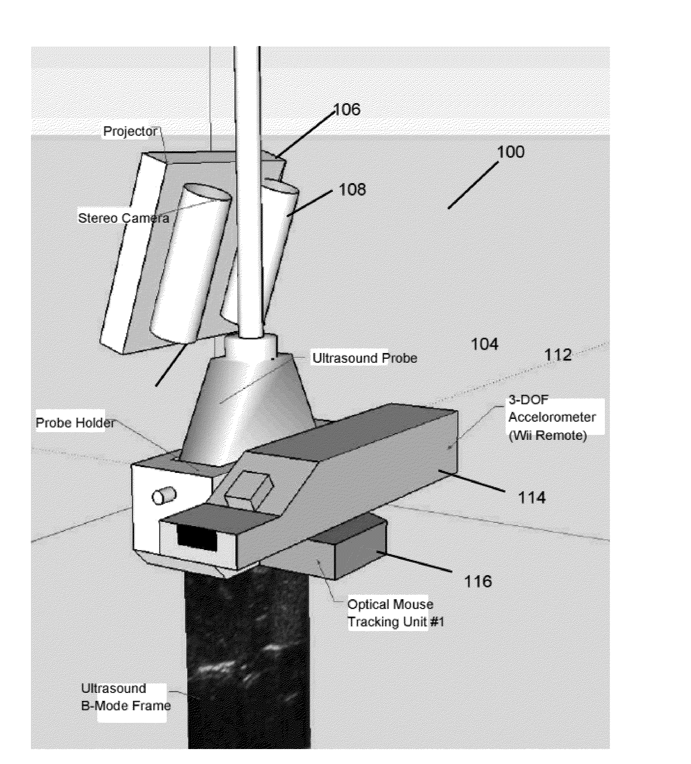
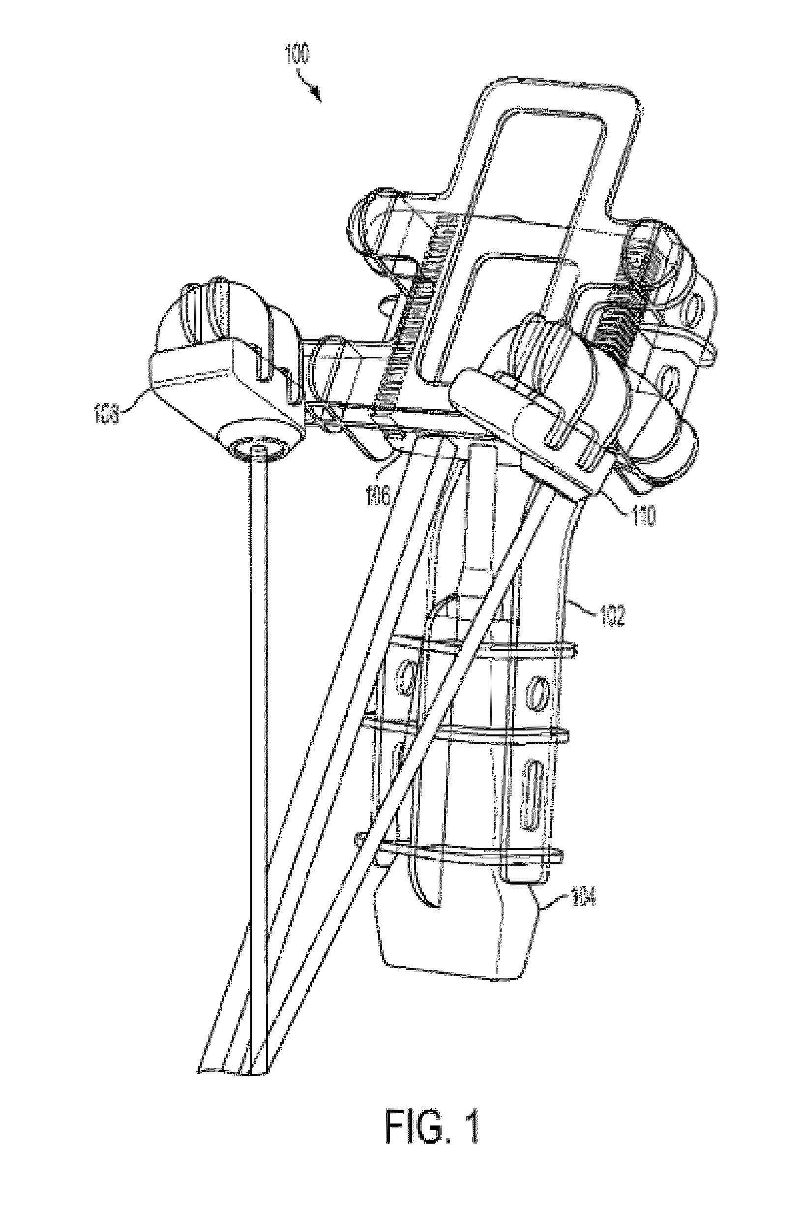
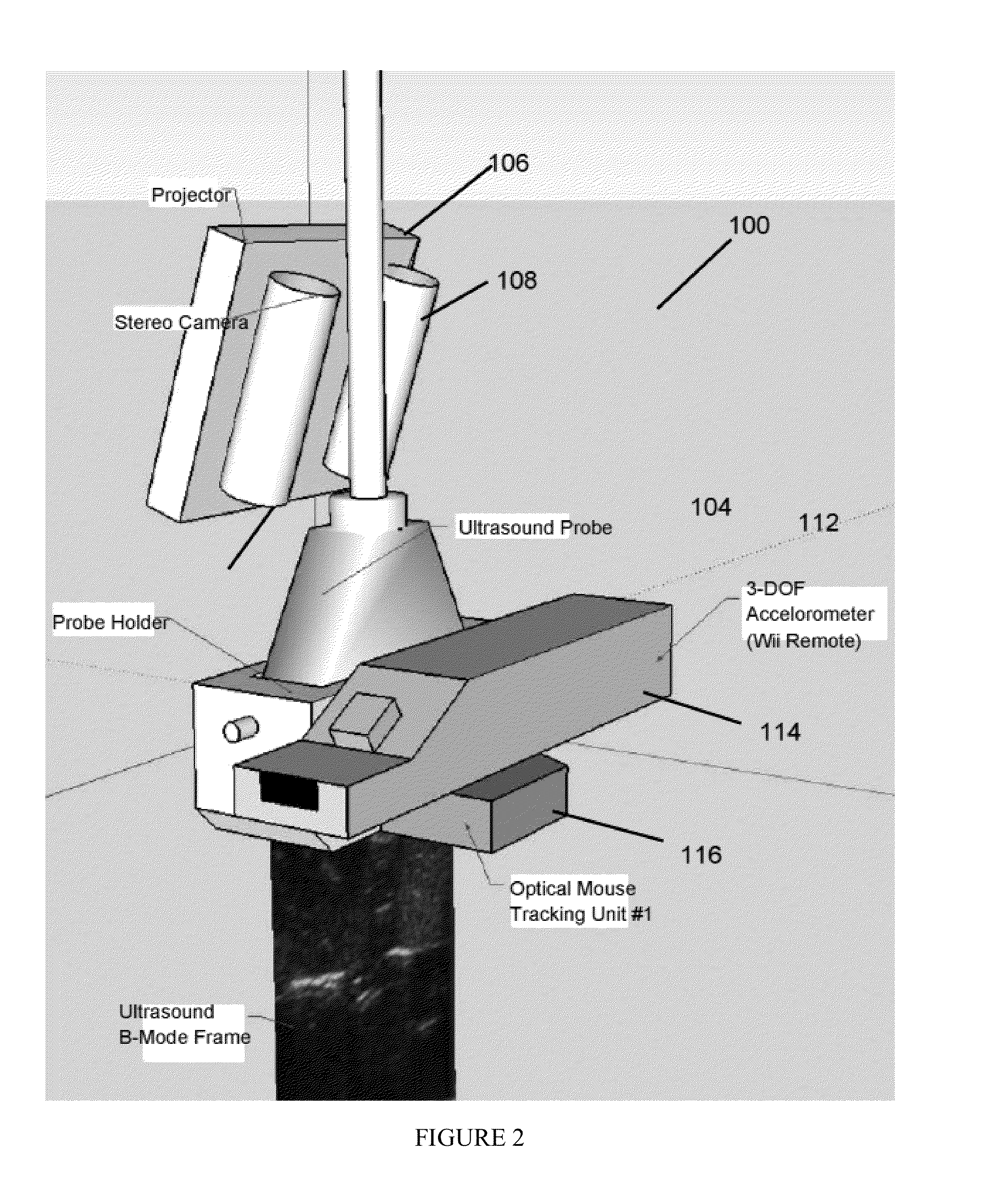
An augmentation device for an imaging system has a bracket structured to be attachable to an imaging component, and a projector attached to the bracket. The projector is arranged and configured to project an image onto a surface in conjunction with imaging by the imaging system. A system for image-guided surgery has an imaging system, and a projector configured to project an image or pattern onto a region of interest during imaging by the imaging system. A capsule imaging device has an imaging system, and a local sensor system. The local sensor system provides information to reconstruct positions of the capsule endoscope free from external monitoring equipment.

Description

CROSS-REFERENCE OF RELATED APPLICATION[0001]This application claims priority to U.S. Provisional Application No. US61 / 545,168 filed Oct. 9, 2011, U.S. Provisional Application No. US61 / 603,625, filed Feb. 27, 2012, and U.S. Provisional Application No. US61 / 657,441, filed Jun. 8, 2012, the entire contents of which are hereby incorporated by reference.BACKGROUND[0002]1. Field of Invention[0003]The field of the currently claimed embodiments of this invention relate to imaging devices and to augmentation devices for these imaging devices, and more particularly to such devices that have one or more of a camera, one or more of a projector, and / or a set of local sensors for observation and imaging of, projecting onto, and tracking within and around a region of interest.[0004]2. Discussion of Related Art[0005]Image-guided surgery (IGS) can be defined as a surgical or intervention procedure where the doctor uses indirect visualization to operate, i.e. by employing imaging instruments in real ...

Claims


Application Information

Patent Timeline
IPC IPC(8): A61B19/00A61B6/00A61B8/08A61B8/00A61B19/08A61B5/06A61B5/055A61B5/00A61B6/03
CPCA61B6/12A61B8/587A61B8/5261A61B2019/5227A61B2019/524A61B2019/5276A61B6/4417A61B19/088A61B8/488A61B8/4416A61B6/4441A61B6/032A61B19/5244A61B5/062A61B5/055A61B5/0077A61B5/0075A61B5/742A61B19/08A61B19/56A61B2017/00707A61B2017/00725A61B2019/5248A61B2019/5263A61B2019/5291A61B2019/5293A61B2019/085A61B8/0841A61B8/4254A61B2034/2048A61B2034/2063A61B2090/366A61B34/20A61B34/25A61B46/00A61B2046/205A61B46/40A61B2090/371A61B2090/3762A61B2090/378A61B2090/365
Inventor BOCTOR, EMAD MIKHAILHAGER, GREGORY DONALDSTOLKA, PHILIPP JAKOBHEISENBERG, DOROTHEE
Owner CLEAR GUIDE MEDICAL
Learn More