
Covering for a medical scoping device

a medical and scope technology, applied in the field of endoscopic examinations/procedures, can solve the problems of affecting the visualisation effect, affecting the patient's experience, and affecting the patient's experience, and achieve the effect of further enhancing the visualisation

Active Publication Date: 2013-04-11
KEYMED MEDICAL & INDAL EQUIP
View PDF8 Cites 69 Cited by
  • Summary
  • Abstract
  • Description
  • Claims
  • Application Information

AI Technical Summary

Benefits of technology

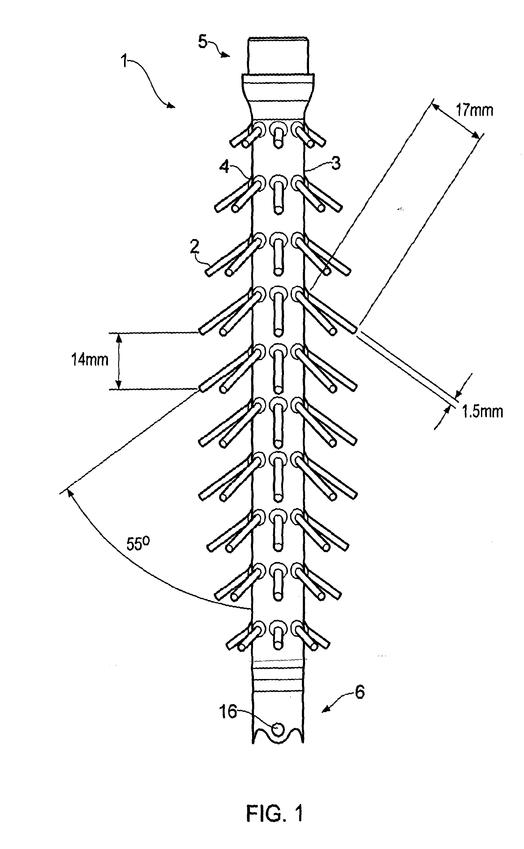
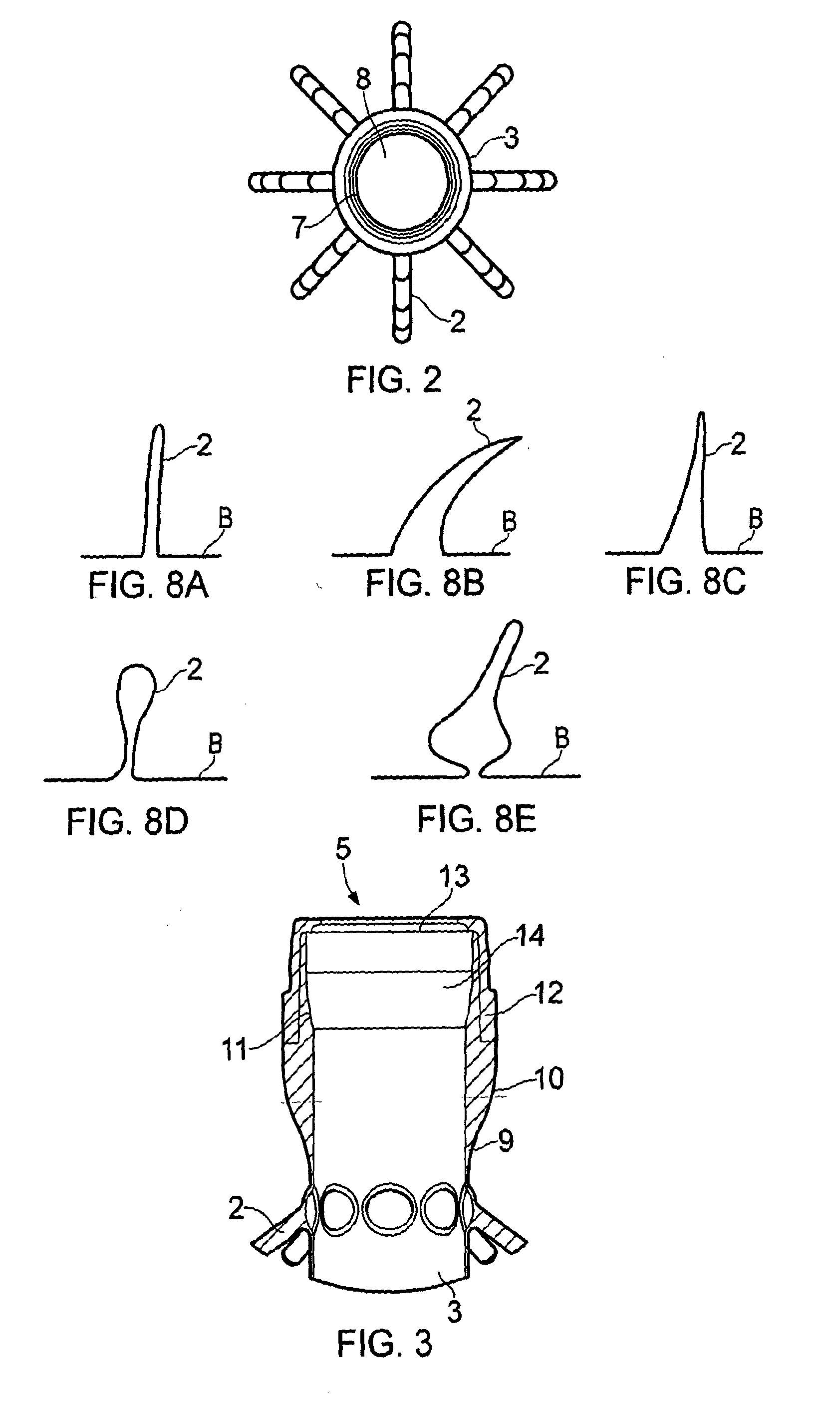
The invention provides a medical scoping device with a cover that can be used to remove air from a patient's bowel or small intestine during a medical examination. The cover has a unique design that can hold the device in place and expand the body passage wall to improve visualisation. The device also has projecting elements that can hold the tip in a central position to prevent flipping back and maintain position during therapy. Overall, the invention improves the efficiency and accuracy of medical examinations and procedures.

Problems solved by technology

Whilst colonoscopic and enteroscopic examinations are the most effective techniques to assess the state of health of the bowel, they are inconvenient, uncomfortable, expensive procedures that are associated with significant risks of potentially serious complications.
A further disadvantage of colonoscopic and enteroscopic procedures is that they are time consuming for patients and medical personnel alike, the procedure can take anywhere from 20 minutes to 2 hours depending on how difficult it is to advance a scope through the colon or small intestine.
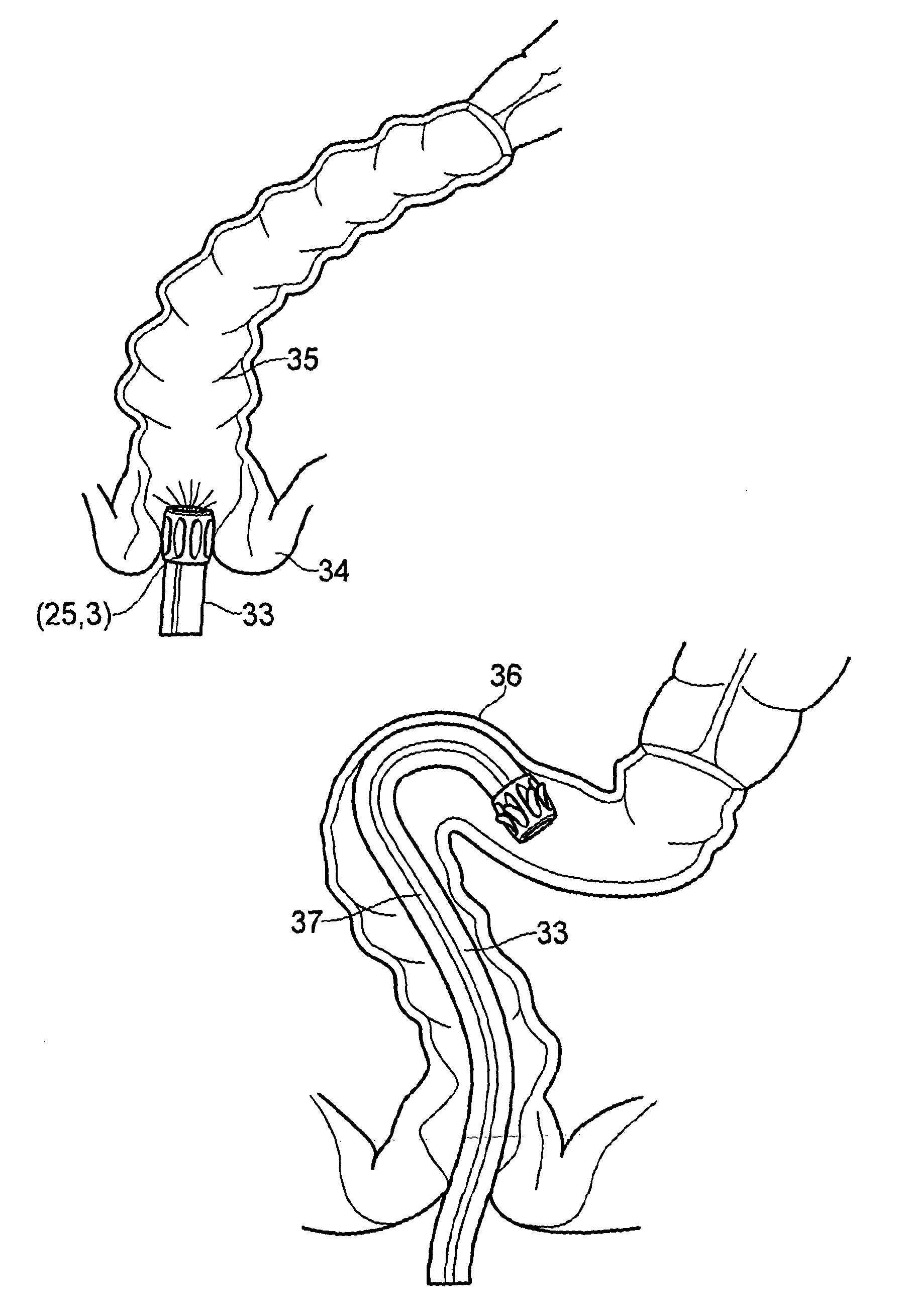
Firstly, the anatomy of the colon is such that the lining is thrown into folds. As the tip of the endoscope passes along the lumen of the colon, these folds hamper the endoscopist's ability to visualise the entire surface of the mucosa and in particular, detect pre-malignant and malignant lesions tucked away on the proximal face of these folds during extubation.
Secondly, the position of the tip of may be difficult to maintain from the moment at which a lesion or polyp is detected to the completion of any therapeutic procedure. As the colonoscope is withdrawn the tip does not travel back at a constant speed but rather with jerks and slippages particularly when traversing a bend or length of colon where the bowel has been concertinaed over the endoscope shaft during intubation. The tip of the device may, at any moment, slip backwards thereby causing the clinician to lose position. If tip position is lost, the clinician is required to relocate the lesion or polyp for the therapeutic procedure to be continued.
The colonoscopic procedure is not simple because the bowel is long and convoluted.
If the stretching is continued or severe while the endoscopist pushes round the bend, the patient experiences pain the blood pressure falls and the pulse slows.
Loop formation is the main cause of failure or delay in completing an examination.
It is responsible for the pain experienced by the patient and the need for heavy sedation that in turn leads to cardio-respiratory complications.
It is also the major cause of perforation in patients not undergoing a therapeutic procedure.
In practice however little improvement is achieved in overcoming looping.
The double balloon enteroscope requires a substantial amount of additional kit, a high level of operator skill in timing the sequential inflation and deflation of the balloons and moreover it is a lengthy procedure sometimes taking hours.
The Aer-O-Scope™ provides low pressure colon insufflations with CO2 to propel the balloon along “slippery” colon walls without forceful maneuvering but cannot be used for biopsy or therapy.
Newer methods for the detection of polyps and cancer using non-invasive technology may be identified but to obtain biopsies, remove polyps and to treat intra-colonic lesions no alternatives have appeared to date.

Method used


Image

  • Covering for a medical scoping device
  • Covering for a medical scoping device
  • Covering for a medical scoping device

Examples


Embodiment Construction

[0038]Throughout the description and claims of this specification, the words “comprise” and “contain” and variations of them mean “including but not limited to”, and they are not intended to (and do not) exclude other moieties, additives, components, integers or steps. Throughout the description and claims of this specification, the singular encompasses the plural unless the context otherwise requires. In particular, where the indefinite article is used, the specification is to be understood as contemplating plurality as well as singularity, unless the context requires otherwise.

[0039]Features, integers, characteristics, compounds, chemical moieties or groups described in conjunction with a particular aspect, embodiment or example of the invention are to be understood to be applicable to any other aspect, embodiment or example described herein unless incompatible therewith. All of the features disclosed in this specification (including any accompanying claims, abstract and drawings)...


PUM


Abstract

The present invention relates to a cover having a plurality of moveable, external, angled projecting elements for use with flexible medical scoping devices such as endoscopesor enteroscopes. The invention includes the cover with an over cuff and use of the disposable removable covering in methods of medical scoping procedures or examinations. The invention also includes an applicator for assisting in placing the covering about or over a medical device and a kit of parts.

Description

[0001]The present invention relates to a covering or sheath or sleeve or cuff having external projections for use with a medical device and in particular for use with flexible medical scoping devices such as endoscopes or enteroscopes. The invention includes inter alia use of the disposable removable covering in methods of medical scoping procedures or examinations, particularly but not exclusively, where the site is the colon or small intestine. The invention also includes a kit including an applicator for assisting in placing the covering about or over a medical scoping device.BACKGROUND[0002]In endoscopic examinations / procedures, flexible instruments designed to view the gastro-intestinal tract are inserted along a body cavity to an internal part such as the stomach, duodenum, small intestine or large intestine. The instruments are provided with fibre-optic or charge-couple device (CCD) cameras which enable images to be transmitted around bends and images to be produced to displa...

Claims


Application Information

Patent Timeline
IPC IPC(8): A61B1/00A61B1/015
CPCA61B1/00075A61B1/0008A61B1/00135A61B1/00142A61B1/015A61B1/31A61B1/32A61B1/00137A61B1/00147A61B1/00154A61B1/01A61M25/04A61M29/00A61B1/00089A61B1/00148A61B1/00101A61B1/00A61M2025/006
Inventor AXON, ANTHONY
Owner KEYMED MEDICAL & INDAL EQUIP
Learn More