Highly sensitive method for the detection of cytosine methylation patterns
a methylation pattern and high-sensitivity technology, applied in the field of cytosine methylation detection in dna samples, can solve the problems of inability to investigate cells for thousands of possible methylation analyses, inability to reliably analyze very small fragments of small sample quantities, and complete loss of epigenetic information, which is carried by 5-methylcytosines
- Summary
- Abstract
- Description
- Claims
- Application Information
AI Technical Summary
Benefits of technology
Problems solved by technology
Method used
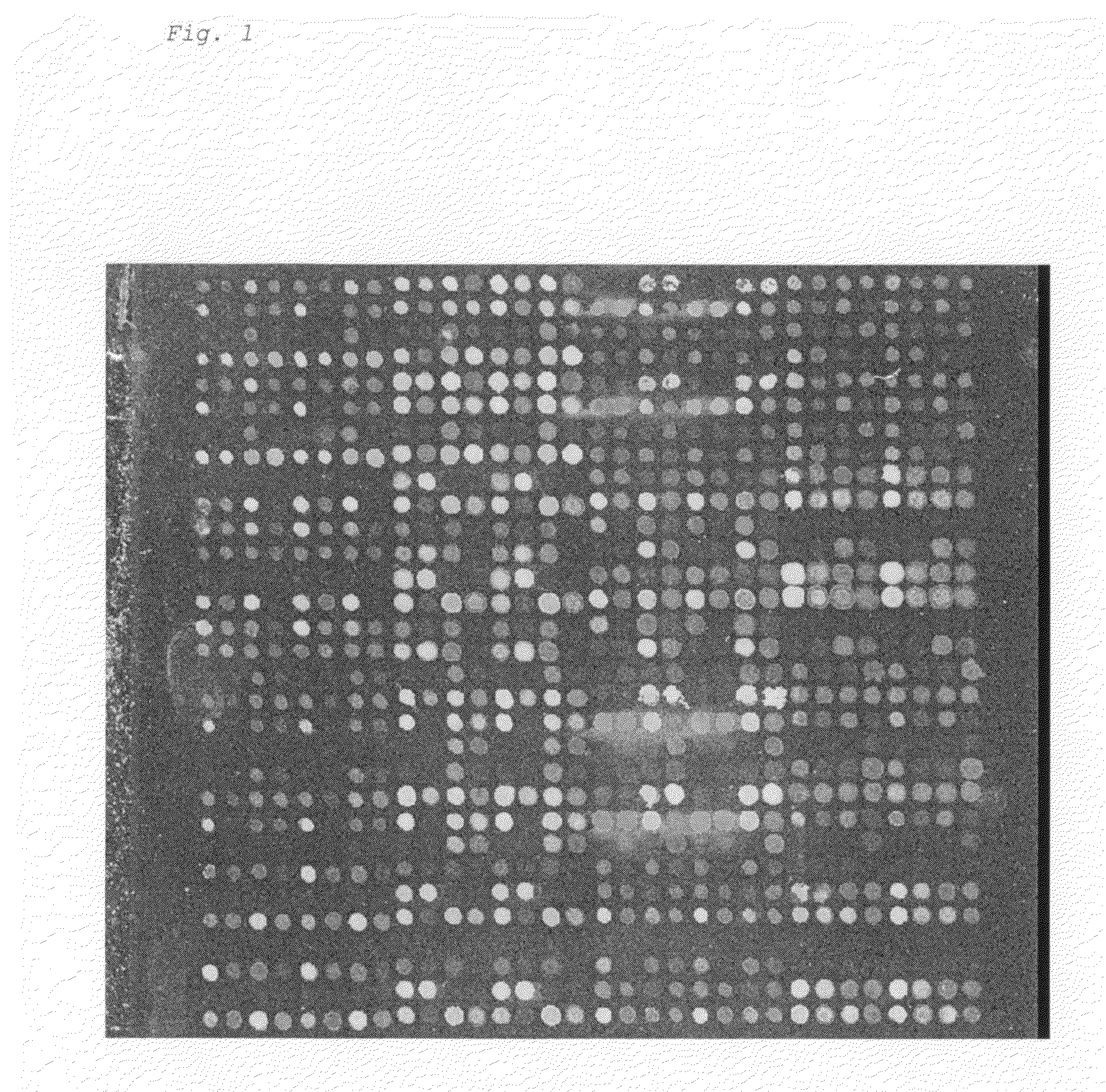
Image

Examples
example 1
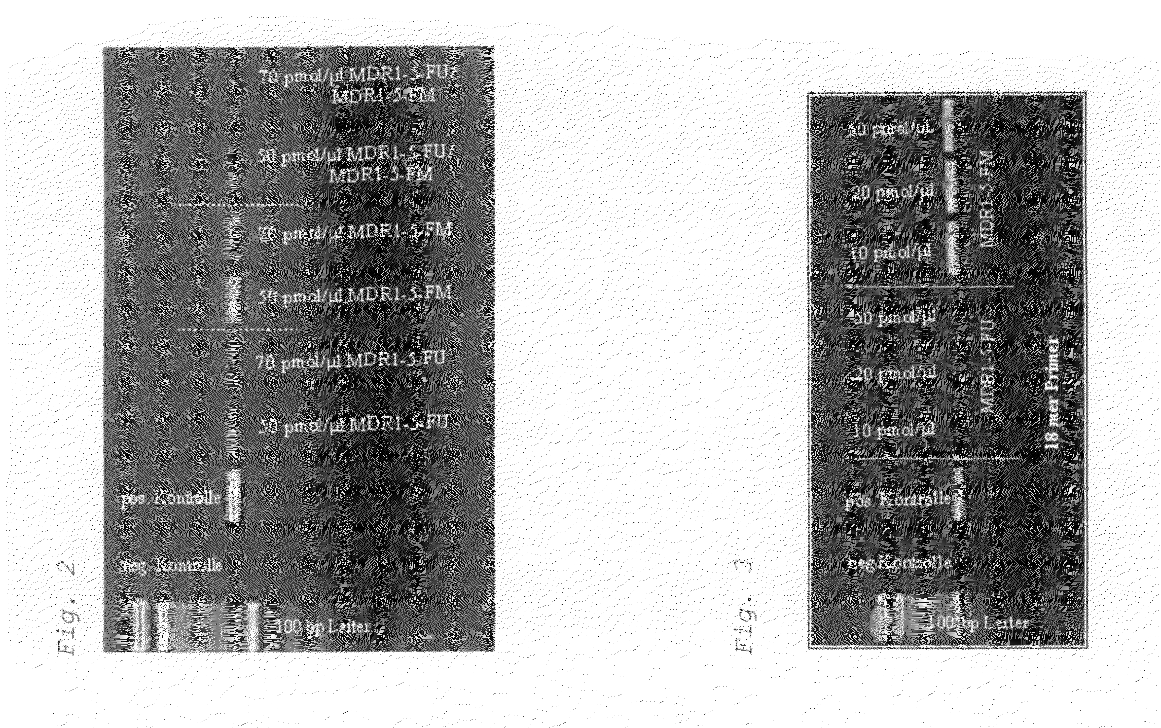
Methylation-Sensitive Amplification of the MDR1 Gene with the Use of PNA Blocking Probes (PCR Clamping) in the MDR-1 Gene
[0123]a) Methylation-Specific PCR with PNA Probes (PNA Blocking Probes), Whose Binding Sites do not Overlap with Those of the Primers.
[0124]First, PCR conditions were defined for bisulfite-treated DNA, by which an allele-specific influence of the PNA blocking probe on the PCR can be recognized. In this first experiment, the binding sites between PNA and the primers do not overlap.
[0125]First, the influence of an 10-mer PNA of the sequence AAAATGTGTT (SEQ ID NO:1) was tested in a PCR with the primers TAAGTATGTTGAAGAAAGATTATTGTAG (SEQ ID NO: 2) and TAAAAACTATCCCATAATAACTCCCAAC (SEQ ID NO: 3). An effect of the PNA on the PCR reaction cannot be measured under standard PCR conditions with an annealing temperature of 55° C. The standard cycler program used for the amplication of the MDR-1 fragment uses the following program steps:
Step 1:T = 96° C. 20 minStep 2:T = 96° ...
example 2
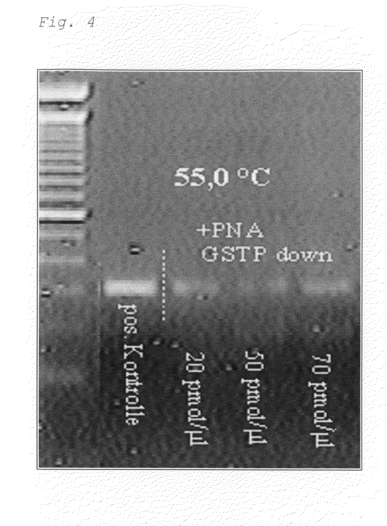
Methylation-Sensitive Amplification of a Fragment of the GSTpi Gene
[0138]Specific CpG positions of the GSTPi gene were identified as tumor markers for prostate cancer. In a pair of primers selected for the experiments, GGAAAGAGGGAAAGGTTTT (SEQ ID NO: 9) and TACTAAAAACTCTAAACCCCAT (SEQ ID NO: 10), a primer is localized such that it precisely bounds the PNA sequence CCCCGAAAACGCG (SEQ ID NO: 11) (or CCCTGAAAATGTG (SEQ ID NO: 12)). The PNA sequence contains three relevant CpG positions now in distinction to those PNAs used for the MDR1 fragment. The relevant CpGs of the GSTPi fragments are present in “normal” DNA unmethylated, i.e., DNA not originating from tumor patients. The PNA “GSTP-down” of the sequence CCCTGAAAATGTG (SEQ ID NO: 12) which is given for the reaction batch should thus have a recognizable influence on the PCR reaction, but not the corresponding PNA “GSTP-up” CCCCGAAAACGCG (SEQ ID NO: 11).
[0139]The PNA “GSTP-down” was added in three different concentrations to the test...
example 3
Different Possibilities for the Use of Probes Suppressing the PCR in a Methylation-Specific Manner on the Example of the GSTPi Gene
[0146]FIG. 6 shows several possibilities of how to arrange the primers in the sense of a methylation-sensitive application for a given template sequence. For explanation, FIG. 6a shows the templates present after the bisulfite treatment: DNA 1 corresponding to an originally methylated DNA sample and DNA 2 corresponding to an originally unmethylated DNA sample.
[0147]FIG. 6b shows the arrangement of one of the primers in the sense of an allele-specific PCR or a methylation-specific PCR (MSP). In this case, only the amplification of the methylated DNA 1 could occur with the use of the primer shown.
[0148]FIGS. 6c and 6d show how correspondingly unmethylation-specific primers can be used, either by the use of degenerated positions (6c) or universal bases (here, inosine) in FIG. 6d.
[0149]Several possibilities are presented in FIG. 7, of how the primers and pr...
PUM
| Property | Measurement | Unit |
|---|---|---|
| temperature | aaaaa | aaaaa |
| temperature | aaaaa | aaaaa |
| temperatures | aaaaa | aaaaa |
Abstract
Description
Claims
Application Information
 - R&D
- Intellectual Property
- Life Sciences
- Materials
- Tech Scout
- Unparalleled Data Quality
- Higher Quality Content
- 60% Fewer Hallucinations
Browse by: Latest US Patents, China's latest patents, Technical Efficacy Thesaurus, Application Domain, Technology Topic, Popular Technical Reports.
© 2025 PatSnap. All rights reserved.Legal|Privacy policy|Modern Slavery Act Transparency Statement|Sitemap|About US| Contact US: help@patsnap.com