
Method and system for intrabody imaging

a technology for intrabody imaging and catheters, applied in the field of medical devices, can solve the problems of difficult use of above covers and discomfort of patients whose lumens are being probed, and achieve the effect of increasing wave conductivity and substantially constant flexibility coefficient of the walls of the adjustable chamber

Active Publication Date: 2012-04-12
COGENTIX MEDICAL
View PDF5 Cites 16 Cited by
  • Summary
  • Abstract
  • Description
  • Claims
  • Application Information

AI Technical Summary

Benefits of technology

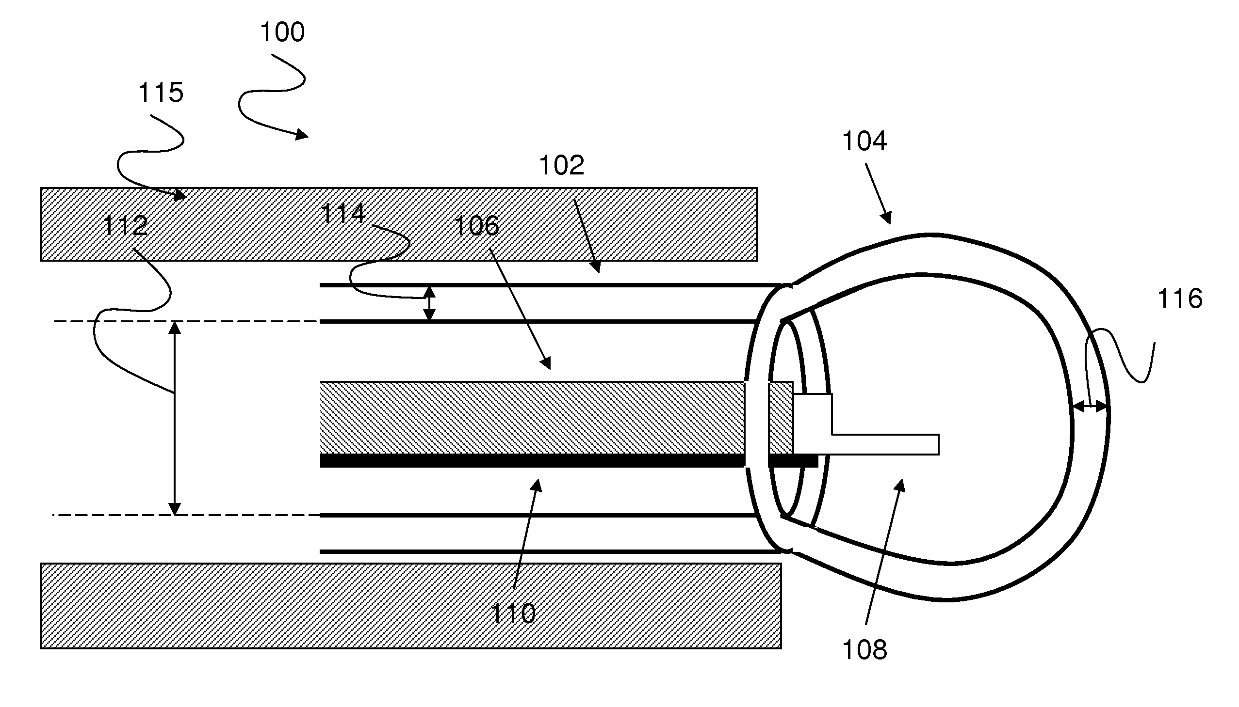
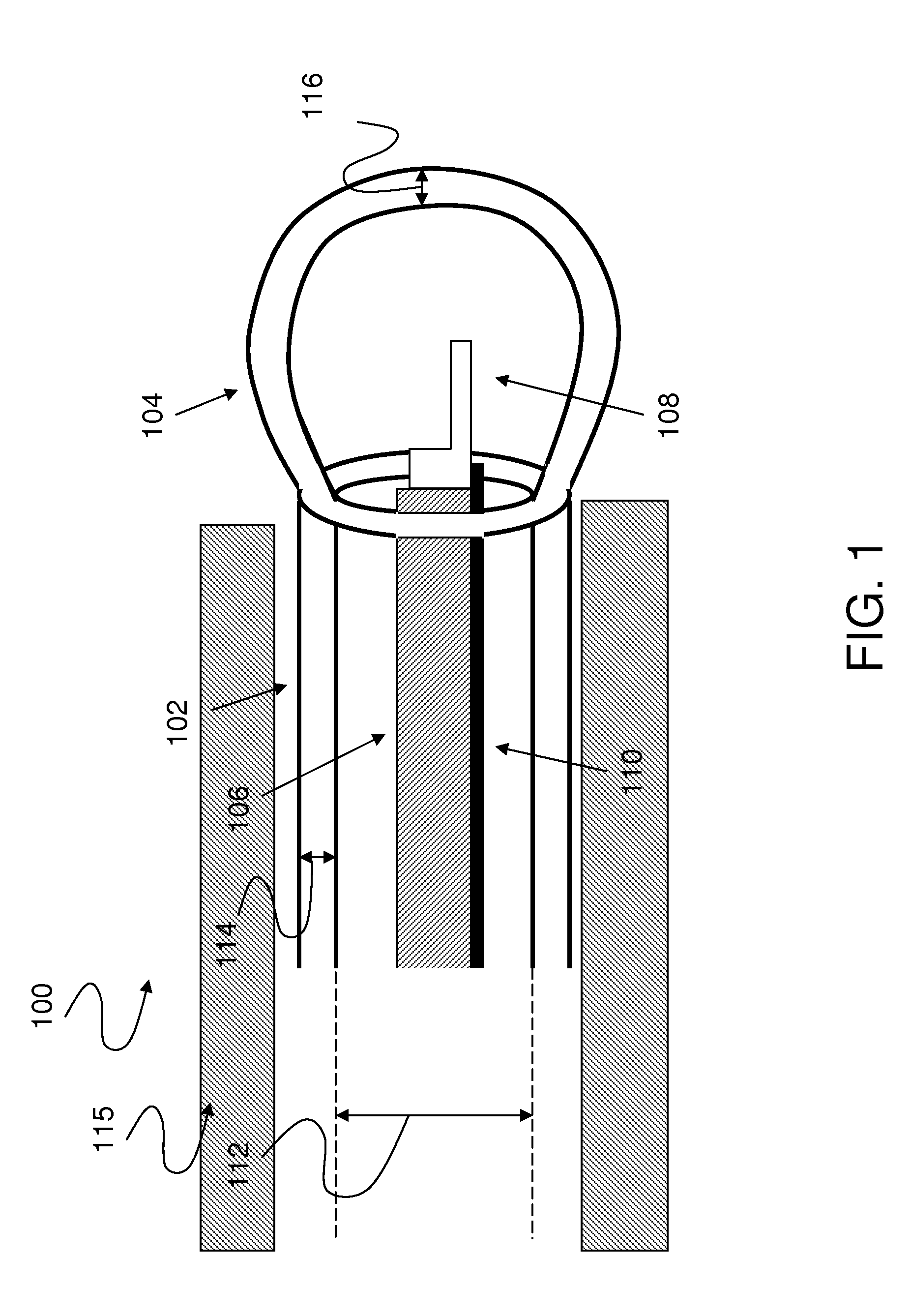
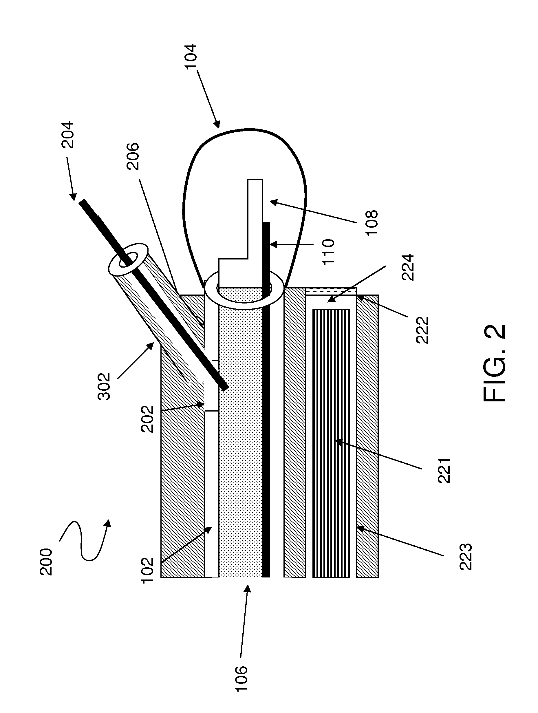
[0009]The present invention, in some embodiments thereof, relates to increasing wave conductivity to allow imaging of a targeted anatomic site in a body by an imager, such as an ultrasound transducer which is connected to the distal end of an intrabody guiding tube, such as a catheter which is designed to pass in a working channel of an endoscope. The channel, through which the ultrasound transducer is passed, is covered with an adjustable chamber, which may be filled with an ultrasound conductive medium while inside the body lumen. The flexibility coefficient of the walls of the adjustable chamber may be substantially constant. The adjustable chamber may fit a working channel of an endoscope. The filling adjusts the volume of the adjustable chamber to match the size and shape of the body lumen.

Problems solved by technology

Such a procedure, however, may cause discomfort to the patient whose lumen is being probed.
Thus, the above cover may be difficult to use.

Method used


Image

  • Method and system for intrabody imaging
  • Method and system for intrabody imaging
  • Method and system for intrabody imaging

Examples


Embodiment Construction

[0039]The present invention, in some embodiments thereof, relates to medical devices and, more particularly, but not exclusively, to catheters, endoscopes, endoscopic tools, and intrabody probes. Some embodiments of the present invention relate to a medical sonography procedure in which an endoscope is used for conveying an ultrasound transducer via a body lumen toward a targeted anatomic site.

[0040]An aspect of some embodiments of the present invention relates to a device for intrabody guiding, optionally disposable, that includes a catheter with a shape adjustable chamber, referred to herein as an adjustable chamber, that is placed at the distal end thereof and covers at least a distal end of an imager, such as an ultrasound catheter.

[0041]The adjustable chamber is designed to be filled with a wave conductive medium, such as an ultrasound conductive medium, that increases wave conductivity in the space between the ultrasound imager and a body tissue in proximity to a targeted anat...


PUM


Abstract

A catheter that comprises a catheter configured for housing at least a portion of a catheter configured for insertion into a body lumen in proximity of a targeted anatomical site and having an imager at a distal end thereof and an adjustable chamber configured for covering the imager. The catheter is configured for introducing a wave conductive medium to the adjustable chamber to increase wave conductivity between said targeted anatomical site and the imager.

Description

RELATED APPLICATIONS[0001]This application is a continuation-in-part (CIP) of U.S. patent application Ser. No. 12 / 486,795 filed Jun. 18, 2009, which claims the benefit of priority under 35 USC 119(e) of U.S. Provisional Patent Application No. 61 / 129,344 filed Jun. 19, 2008.[0002]The contents of the above applications are incorporated herein by reference in their entirety.FIELD AND BACKGROUND OF THE INVENTION[0003]The present invention, in some embodiments thereof, relates to medical devices and, more particularly, but not exclusively, to catheters, endoscopes, endoscopic tools, and intrabody probes.[0004]Endoscopy is a minimally invasive diagnostic medical procedure that is used to assess the interior surfaces of an organ or of a body by inserting an insertion tube of an endoscope into a body lumen, or cavity, of a patient. A typical endoscope includes a rigid or flexible endoscopic insertion tube and an endoscope control unit, such as a handle, for allowing a user to hold and / or co...

Claims


Application Information

Patent Timeline
IPC IPC(8): A61B8/14
CPCA61B1/00098A61B1/018A61B8/445A61B8/14A61B8/58A61B8/12
Inventor HADANI, RONLANDMAN, MARK
Owner COGENTIX MEDICAL
Learn More