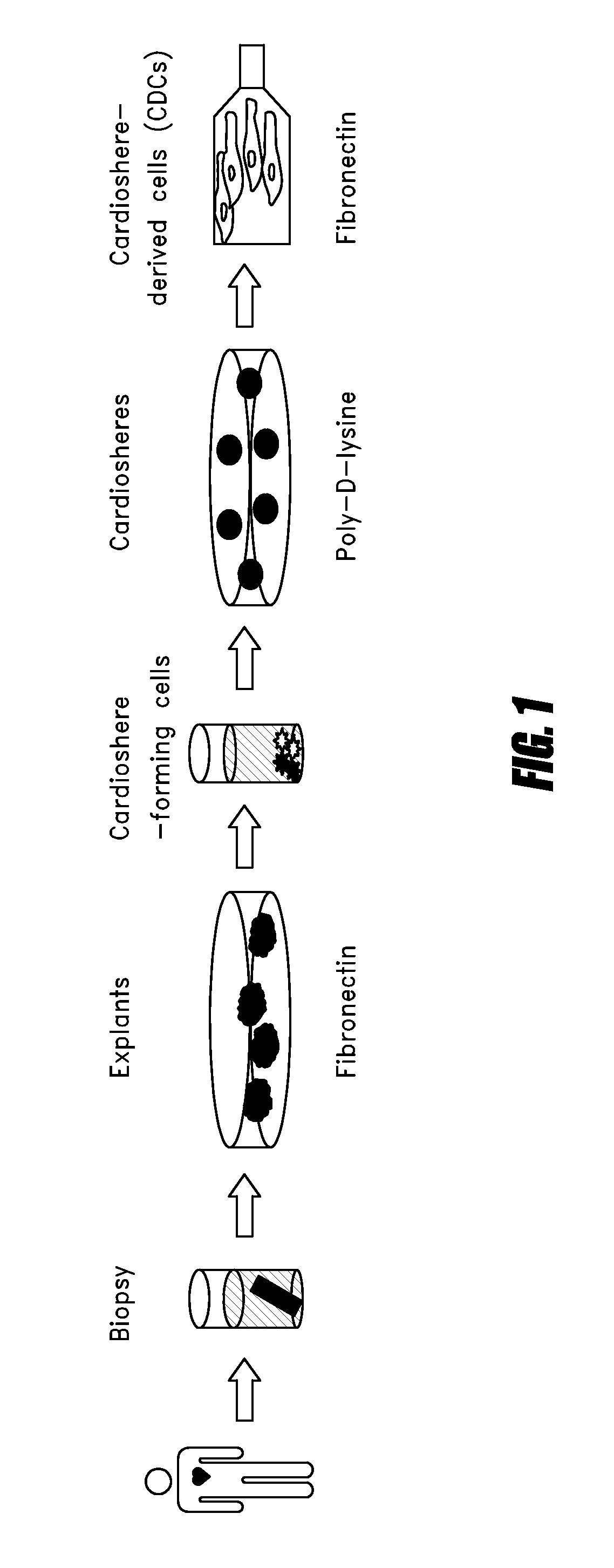
Systems and methods for cardiac tissue repair
a tissue repair and cardiac tissue technology, applied in the field of systems and methods for cardiac tissue repair, can solve the problems of reducing the quality of life of patients, reducing the ability of coronary heart disease to deteriorate into heart failure, and forming teratomas, so as to maximize the migration of cells and increase the viscosity of the matrix
- Summary
- Abstract
- Description
- Claims
- Application Information
AI Technical Summary
Benefits of technology
Problems solved by technology
Method used
Image
Examples
example 1
Matrix Preparation & Cell Survival
[0099]Experiments to test the ability of cells to survive in biomaterials in culture were performed using hyaluronan-based hydrogel. Fibrin, alginate or other biomaterials may be used in addition to or instead of hyaluronan. Thiolated hyaluronan and polyethylene glycol diacrylate (PEGDA, a thiol-reactive crosslinker) were obtained as lyophilized solids. Warm (37° C.) degassed, deionized water was used to dissolve each component separately. Dissolution of the hyaluronan required about 30 minutes of rocking or shaking, while the PEDGA was readily solublized. Once reconstituted, the gel components were diluted in phosphate-buffered saline (pH=7.4). Cardiospheres, CDCs, cardiac stem cells, or mixtures thereof can be resuspended in any solution, and mixed with the hyaluronan solution. Hyaluronan (with or without cells) and PEGDA are then mixed in a 4:1 ratio to create a hydrogel. Gelation occurs within approximately 20 minutes. Diluting the hyaluronan or...
example 2
CDC Survival at 72 Hours in Hyaluronan Hydrogels With or Without Collagen
[0106]In order to further characterize the survival of cells in various matrices, hyaluronan-based hydrogels were reconstituted as described above. CDCs were incorporated into the hydrogels during the crosslinking process (prior to gelation) and seeded on a 96 well plate. The final aqueous cell solution passed readily through a 30-gauge needle.
[0107]Plates were pre-coated with a thin layer of gel prior to seeding the CDC-hydrogels. This was done to prevent cells from settling out of the gel during the time needed for gelation and coming into contact with the polystyrene tissue culture plates. Gelation occurred within approximately 20 minutes. In several embodiments, a 20 minute gelation time is rapid enough for the hydrogel to improve cell retention within the myocardium yet also slow enough to allow for complete passage of the cell-biomaterial solution to pass fully through the delivery mechanisms (e.g., cathe...
example 3
Migration Assay
[0114]As cells incorporated into a biomaterial likely have to migrate from the biomaterial in order to provide the most efficacious repair or regeneration of damaged tissue, the present experiment was designed to evaluate the in vitro migratory potential of cells incorporated within the hyaluronan and collagen-supplemented hyaluronan hydrogels. While either cardiospheres, CDCs or other cells types may be delivered via a biomaterial, in this experiment, CDCs were labeled with calcein to enable the tracking of migrating cells. Cell labeling was performed by established techniques. Calcein-labeled CDCs were incorporated into various hydrogels at a cell concentration of 10,000 cells / μL. CDCs within the hydrogels were cultured in a transwell plate setup that allowed for cell migration from the upper chamber, through pores in the bottom of the chamber insert, and into the lower chamber where they could be detected. Fetal bovine serum was used as a chemoattractant in the low...
PUM
 Abstract
Description
Claims
Application Information
 - R&D
- Intellectual Property
- Life Sciences
- Materials
- Tech Scout
- Unparalleled Data Quality
- Higher Quality Content
- 60% Fewer Hallucinations
Browse by: Latest US Patents, China's latest patents, Technical Efficacy Thesaurus, Application Domain, Technology Topic, Popular Technical Reports.
© 2025 PatSnap. All rights reserved.Legal|Privacy policy|Modern Slavery Act Transparency Statement|Sitemap|About US| Contact US: help@patsnap.com


