
Method and apparatus for multimodal imaging of biological tissue

a biological tissue and multimodal imaging technology, applied in the field of biological tissue multimodal imaging, can solve the problems of complicated visual tumor localization and precise excision, high cost of treatment, and time-consuming and labor-intensive procedures, and achieve the effect of facilitating real-time tumor delineation and increasing the number of surgical procedures

Inactive Publication Date: 2012-02-02
THE GENERAL HOSPITAL CORP
View PDF4 Cites 13 Cited by
  • Summary
  • Abstract
  • Description
  • Claims
  • Application Information

AI Technical Summary

Benefits of technology

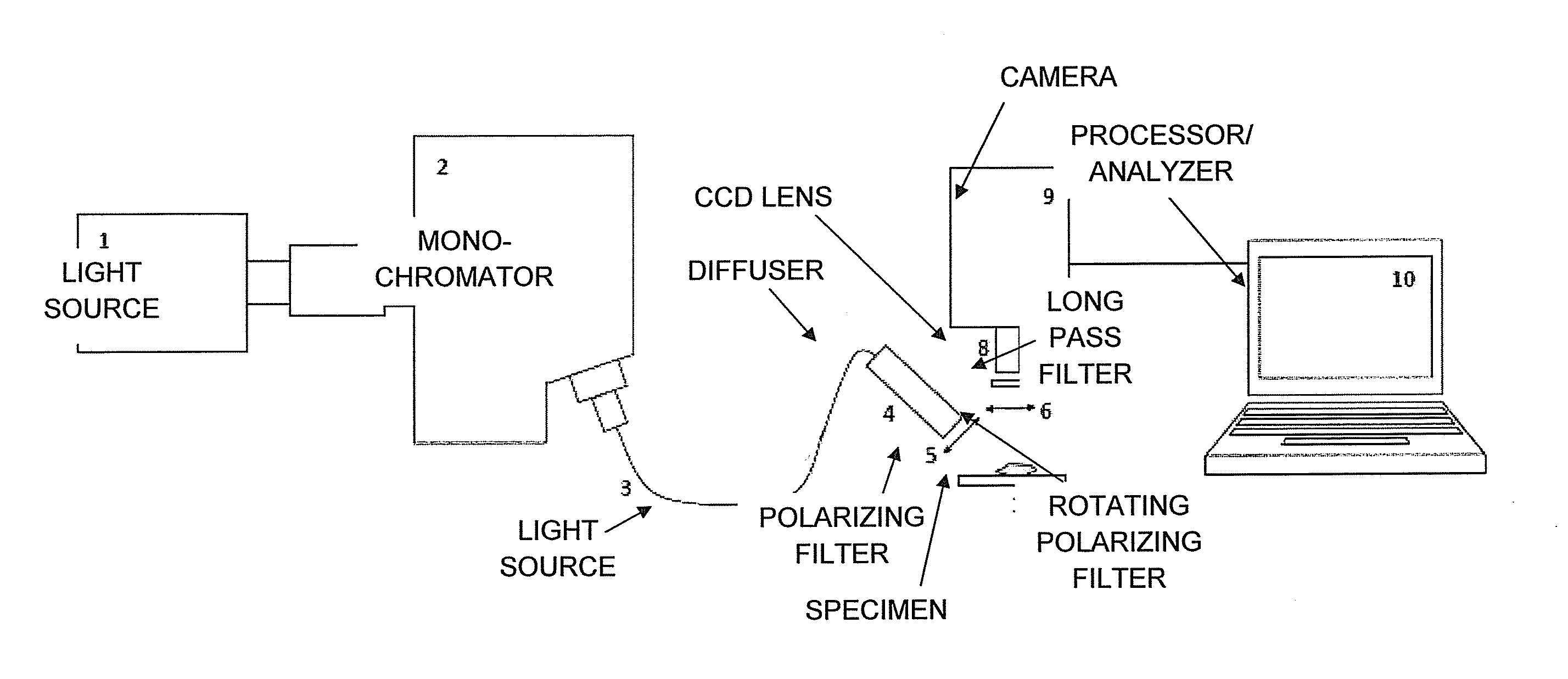
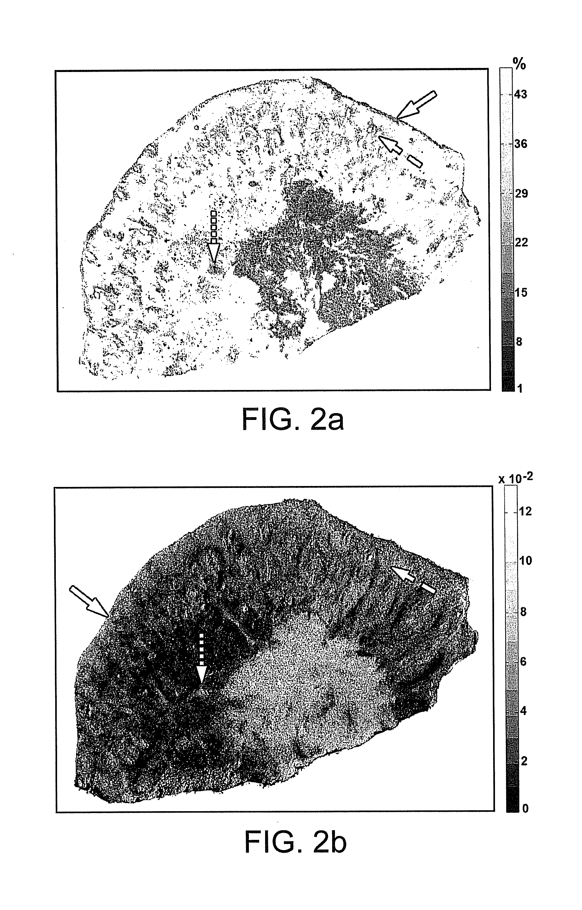
[0010]In one embodiment of the present invention, a tumor demarcation technique can be provided that includes imaging a tissue region or sample treated with a contrast agent such as a dye at two or more particular wavelengths. Imaging light can be provided by monochromatic light sources, or by a broadband light source and two or more monochromatic filters, which can optionally be polarizing filters. A local optical density, reflectance, or other optical property associated with different tissue structures, e.g., various healthy skin structures and cancerous tissue, may exhibit a different variation with respect to wavelength for different structures, even when the values of such properties may be similar for certain tissue types or structures. Accordingly, a method and apparatus for image analysis of tissue using this technique can provide a quick spectroscopic analysis at as few as two (2) interrogation wavelengths that may be sufficient to more reliably distinguish a structure such as cancerous tissue from surrounding healthy tissue. Embodiments of the present invention may therefore provide a method and apparatus that facilitates real-time accurate intraoperative identification and delineation of cancerous tissues or other tissue structures.
[0012]In a further embodiment of the invention, the contrast agent can be methylene blue when in vivo tissue imaging is performed. At least one of the wavelengths used with this contrast agent is preferably between about 600 nm and about 735 nm. For example, a wavelength of about 615 nm and / or about 665 nm may be used, as these wavelengths correspond to absorption maxima of methylene blue and thereby can provide large signals that facilitate identification and / or delineation of different tissue types. When imaging tissue ex vivo, a contrast agent such as toluidine blue can be used.
[0015]In yet another embodiment of the invention, the optical properties determined at two or more wavelengths within the tissue region can be compared to predetermined wavelength-dependent values of such properties for various tissue types. Such comparison of local optical properties at more than one wavelength can provide more accurate identification and delineation of tissue types as compared with measurement of optical properties obtained at a single wavelength. Such predetermined optical properties used for comparison with detected or measured values in a tissue sample can also include derivatives of the measured optical properties with respect to wavelength.
[0017]The methods, system and apparatus provided in accordance with the present invention may be particularly useful for real-time intra-operative imaging and delineation of nonmelanoma skin tumor margins, accelerating the tumor excision process and increasing the number of surgical procedures that can be performed. For example, embodiments of the present invention can be used to guide an actual tumor excision surgery. Such improvements can be provided by incorporating spectral analysis into RFPI procedures and systems that facilitate real-time tumor delineation processes.

Problems solved by technology

However, due to their prevalence, the cost of their treatment exceeds $600 million per year.
However, in many cases the contrast of the lesions is poor and these tumors have poorly defined boundaries, which complicates visual tumor localization and precise excision.
While precise and accurate, MMS is a time-consuming and staff-intensive procedure that generally requires a surgeon trained in dermatopathology, a dedicated laboratory and a technician to prepare and evaluate frozen sections.
However, these techniques generally lack one or more elements necessary for their practical use in a clinical setting.
Even though the MB dye tends to accumulate to a much greater extent in the mitochondria of cancerous cells as compared to normal cells, it is not 100% specific.
However, such techniques may not be able to reliably distinguish between cancerous tissue and certain tissue structures, e.g., hair follicles or the like, that may exhibit similar optical densities.

Method used


Image

  • Method and apparatus for multimodal imaging of biological tissue
  • Method and apparatus for multimodal imaging of biological tissue
  • Method and apparatus for multimodal imaging of biological tissue

Examples


Embodiment Construction

[0028]Biological contrast agents, e.g. stains or dyes such as MB, can distribute differently in different types of tissue. Such variations in dye distribution can lead to differences in spectral absorption and other optical properties, which can depend in part on differences in local dye concentrations. Other factors may also affect the observed optical properties, such as local concentration of blood / hemoglobin or other light-absorbing substances in the tissues. In conventional tissue identification techniques, optical properties of dyed tissue measured using a broadband source or a single wavelength can be compared to identify differences in tissue types based on variation in the dye distribution. However, certain distinct tissue types may exhibit similar optical properties in such measurements, which can impair the identification or differentiation of tissue types in a sample.

[0029]Certain optical properties at two or more particular wavelengths can be measured at a plurality of ...


PUM


Abstract

The present invention is directed to a novel multi-wavelength imaging method and apparatus that enables rapid imaging of tissue regions with accurate identification of tissue types within the region. Optical properties, such as co-polarized or cross-polarized fluorescence or reflectance intensity, optical density and / or reflectance, can be determined at a plurality of locations within the tissue region for each wavelength. Said properties at the two wavelengths, including calculated derivatives of the optical property with respect to wavelength, can be analyzed to image tissue structures and identify tissue types within the tissue region more accurately than can be achieved based on properties measured at a single wavelength.

Description

CROSS REFERENCE TO RELATED APPLICATION[0001]The present application claims priority from U.S. Provisional Patent Application Ser. No. 61 / 169,258 filed Apr. 14, 2009, the disclosure of which is incorporated herein by reference in its entirety.FIELD OF THE INVENTION[0002]The present invention is directed to a method and apparatus that facilitates accurate delineation of cancerous tissue or other particular types of biological tissue in tissue samples by comparing absorbance properties and / or other optical interactions of dyes by different tissue types at two or more particular wavelengths. The method and apparatus can be used to provide, among other things, real-time intraoperative delineation or demarcation of nonmelanoma skin cancers (e.g., basal cell carcinomas).BACKGROUND INFORMATION[0003]Nonmelanoma skin cancers are a very common form of human cancer. Statistically, about one out of four Caucasians will develop at least one lesion during their life time. Basal cell carcinomas (BC...

Claims


Application Information

Patent Timeline
Patent Type & Authority Applications(United States)
IPC IPC(8): A61B6/00
CPCG01N21/6456G01N21/6445
Inventor YAROSLAVSKY, ANNA N.SALOMATINA, ELENAANDERSON, RICHARD R.
Owner THE GENERAL HOSPITAL CORP
Learn More