Image processing apparatus, control method thereof, and program

a technology of image processing and control method, applied in the field of image processing apparatus, can solve the problems of inability to accurately obtain information inside the retina, difficult quantitative evaluation, loss of three-dimensional information,

Inactive Publication Date: 2011-10-06
CANON KK
View PDF4 Cites 20 Cited by
  • Summary
  • Abstract
  • Description
  • Claims
  • Application Information

AI Technical Summary

Problems solved by technology

Although three-dimensional information can be obtained by the observation based on the slit lamp, quantitative evaluation is difficult.
On the other hand, although quantitative evaluation is possible in the fundus picture, three-dimensional information is lost.
However, the information obtained by imaging by the stereoscopic fundus camera is just a combination of images obtained by capturing from different angles, and the information does not include tomographic information inside the retina.
Therefore, there is a problem that the information inside the retina cannot be accurately obtained, as compared to the information of the volumetric images obtained by the OCT.
Therefore, in the conventional OCT volumetric images, it is difficult to present information that allows recognition of the entire image.

Method used


Image

  • Image processing apparatus, control method thereof, and program
  • Image processing apparatus, control method thereof, and program
  • Image processing apparatus, control method thereof, and program

Examples


first embodiment

[0021]Hereinafter, a first embodiment will be described with reference to the drawings. An image processing apparatus according to the present embodiment presents volumetric images that clarify the progression of disease when a plurality of volumetric images indicating temporal changes of glaucoma are obtained by OCT to observe detailed structural changes in the retina. Specifically, information at an initial stage of disease with high approximation accuracy based on OCT volumetric images is used to create a model of sclera with few changes associated with the progression of disease. The model is combined with a plurality of volumetric images after the progression of disease and is displayed. As the created OCT volumetric images are used, the changes in the detailed structure of the retina associated with the progression of disease are clarified by displaying the changes with information of the disc which serves as a comparison index of the changes.

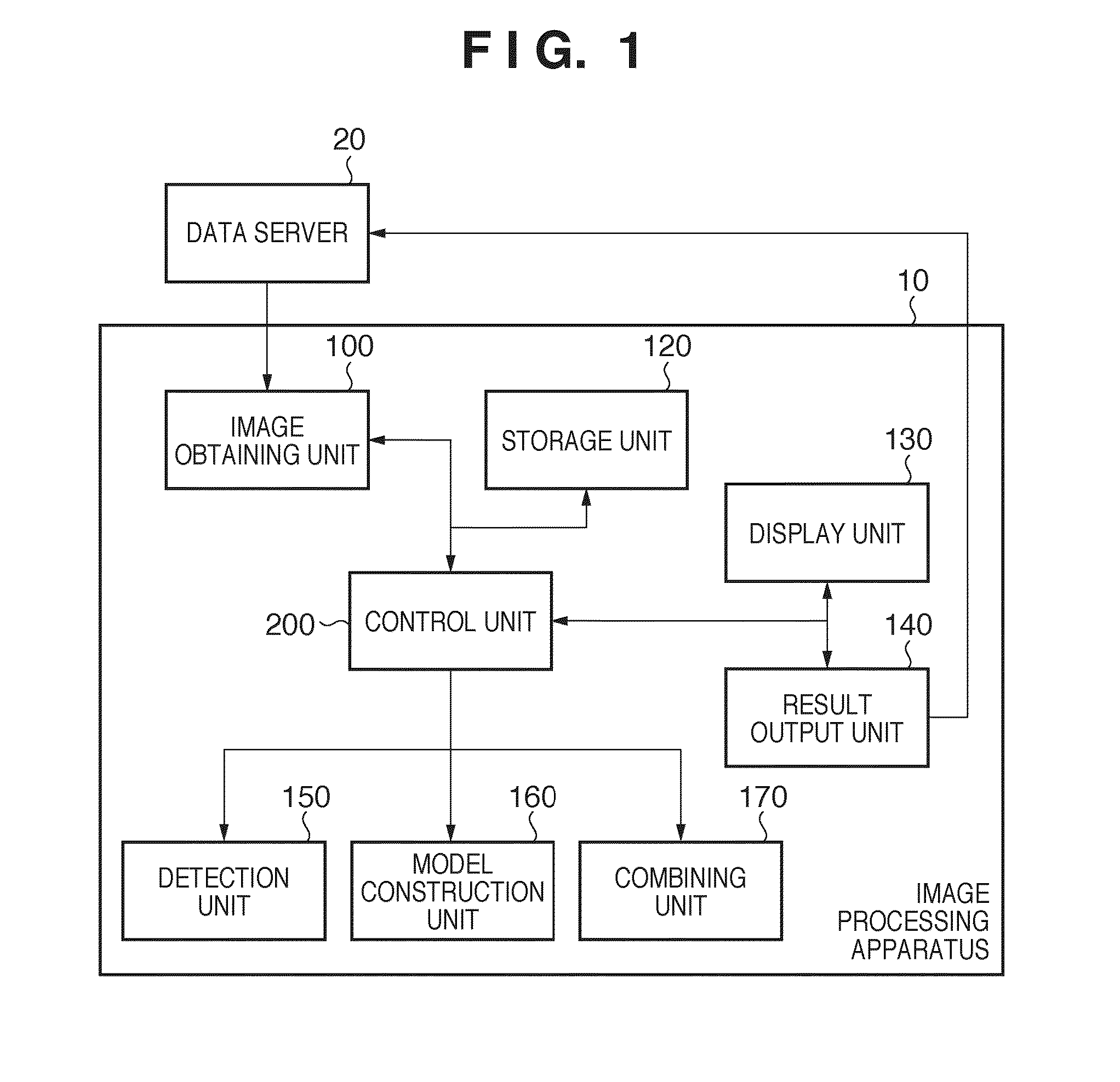
[0022]FIG. 1 is a diagram showing ...

second embodiment

[0047]The information of the RPE boundary and the RPE ends obtained from the OCT volumetric images is used to construct the model of sclera in the first embodiment. However, the RPE ends provide the approximate value of the optic papilla periphery only when there are no findings of peripapillary atrophies (PPA), etc., and it is difficult to construct the model of sclera only from the OCT volumetric images after the progression of disease. In a second embodiment, a case of using image data of an image other than the OCT volumetric images, a fundus image here, to construct a more accurate model to create the model of sclera will be described.

[0048]Hereinafter, a processing procedure of the image processing apparatus 10 of the present embodiment will be described with reference to the flow chart of FIG. 8. Steps S810, S840, and S850 are the same as steps S5210, S240, and S250 of the first embodiment, respectively, and the description will not be repeated. It is assumed that a plurality...


PUM


Abstract

An image processing apparatus including a storage unit configured to store tomographic images in relation to fundus of an examined eye; a detection unit configured to detect a boundary of retinal pigment epithelium and an inner limiting membrane from each of the images and to detect a part where the boundary is discontinuous; a determination unit configured to determine a surface of a sclera model for each of the images by use of the boundary and the inner limiting membrane; a generation unit configured to generate a sclera model including an optic papilla periphery by use of the surface of the sclera model and the part where the boundary is discontinuous; a combining unit configured to combine each of the images and the sclera model to generate a combined image; and a display unit configured to display the combined image generated by the combining unit, is provided.

Description

BACKGROUND OF THE INVENTION[0001]1. Field of the Invention[0002]The present invention relates to an image processing apparatus that captures tomographic images of an observation target, a control method of the image processing apparatus, and a program, and particularly, to a technique applicable to observation of tomograms of an eye portion.[0003]2. Description of the Related Art[0004]In recent years, an apparatus that obtains tomograms of retina by Optical Coherence Tomography (hereinafter, called “OCT”) is implemented in clinical sites of ophthalmology, and new findings are provided. A plurality of tomograms of the retina can be obtained as volumetric images, and changes of each layer associated with the progression of disease can be quantitatively obtained across a wide range. This is expected to lead to highly accurate recognition of the degree of progression or evaluation of the treatment effect.[0005]The relationship with the findings obtained by conventional methods is import...

Claims


Application Information

Patent Timeline
Patent Type & Authority Applications(United States)
IPC IPC(8): G06K9/00
CPCA61B3/102G06T7/0012G06T2207/30041G06T2207/10101G06T7/0083G06T7/12
Inventor YONEZAWA, KEIKOTAKAMA, YASUFUMI
Owner CANON KK
Why Eureka
Try Eureka