
Marker delivery device with obturator

a delivery device and biopsy site technology, applied in the field of medical devices and methods, can solve the problems of affecting the delivery of tissue markers, so as to reduce the risk of clot formation, facilitate the delivery of one or more tissue markers, and improve the delivery

Inactive Publication Date: 2011-07-28
SENORX
View PDF99 Cites 28 Cited by
  • Summary
  • Abstract
  • Description
  • Claims
  • Application Information

AI Technical Summary

Benefits of technology

Minimizes blood backflow and clot formation, enhances the accuracy of marker placement, and facilitates precise localization of biopsy sites during and after procedures, improving surgical planning and execution.

Problems solved by technology

This backflow of blood into the obturator creates a risk of blood clotting.
After the biopsy sample is taken, it may take several days or weeks before the results of the examination of the sample are obtained, and still longer before an appropriate treatment decision is reached.
However, radiographically imageable tissue features, originally detected in a mammogram, may be removed, altered or obscured by the biopsy procedure.

Method used


Image

  • Marker delivery device with obturator
  • Marker delivery device with obturator
  • Marker delivery device with obturator

Examples


Embodiment Construction

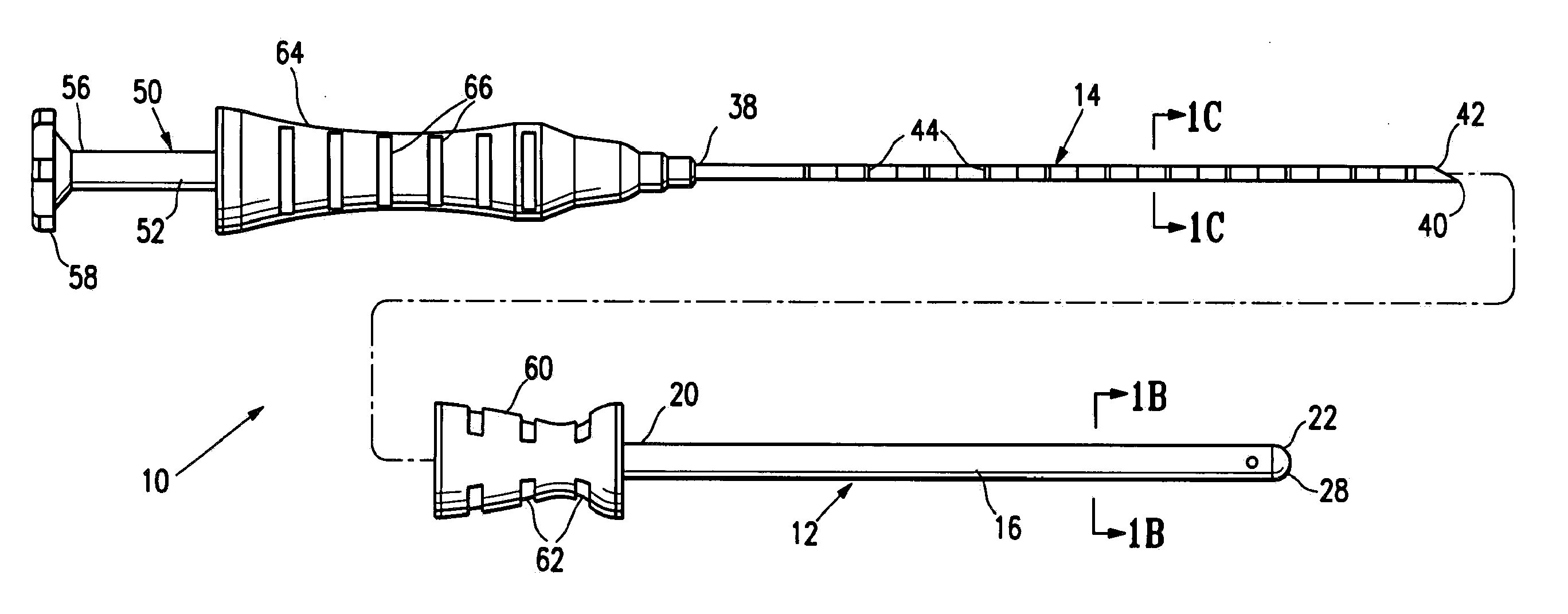
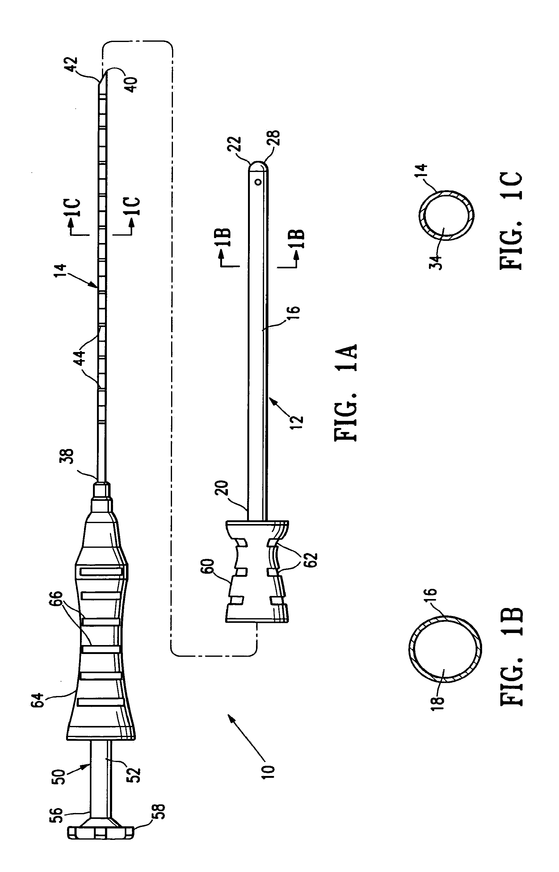
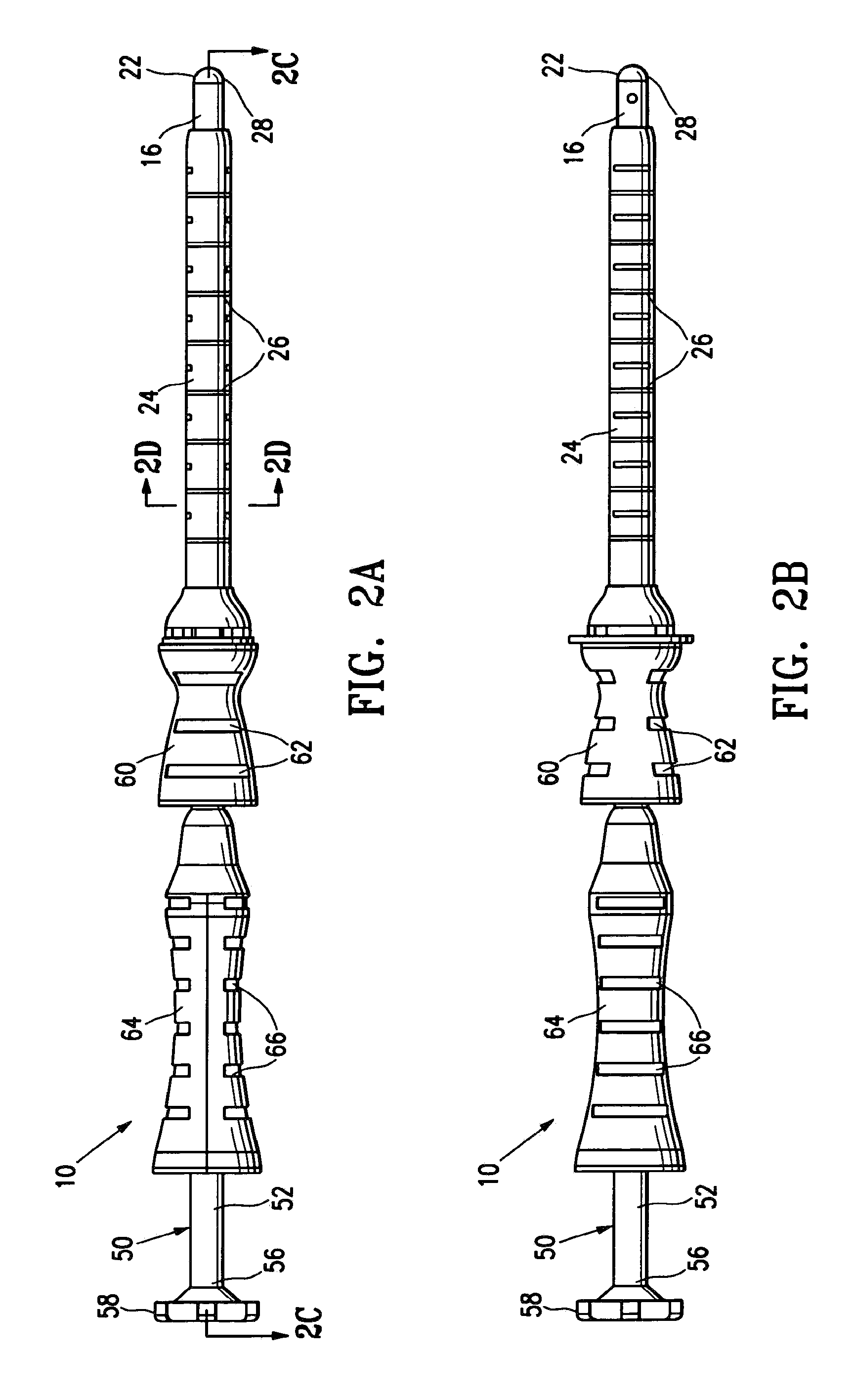
[0045]FIGS. 1A-2D shows an embodiment of a marker delivery device 10 having features of the invention including an obturator 12 and a marker delivery tubular shaft 14. The obturator 12 has an elongated shaft 16, an internal lumen 18, a proximal end 20 and a substantially sealed distal end 22. Preferably, as shown in FIGS. 2A-2D, the obturator 12 is configured to fit within a cannula 24 of a biopsy device, such as SenoRx's EnCor™ Magnetic Resonance Imaging (MRI) Breast Biopsy System. The cannula 24 provides access to the desired location within a patient's body. In some embodiments the cannula 24 includes depth markings 26 which indicate the distance which the obturator 12 has advanced within the patient's body.

[0046]The substantially sealed distal end 22 of the obturator 12 is configured to prevent or minimize the backflow of fluids, such as body fluids, through the internal lumen 18 of the obturator 12. Preferably the substantially sealed distal end 22 is formed of a penetrable mem...


PUM


Abstract

A marker delivery device includes an obturator having an elongated shaft, an internal lumen, a proximal end, and a substantially sealed distal end. The substantially sealed distal end is formed of a penetrable membrane. A marker delivery tube is configured to be slidably disposed within the internal lumen of the obturator. The marker delivery tube has a marker delivery lumen, a proximal end, and a distal tip. The marker delivery lumen is configured to contain one or more tissue markers. The distal tip is configured to puncture the penetrable membrane of the substantially sealed distal end of the obturator to form a passage through which the distal tip extends to facilitate delivery of the one or more tissue markers.

Description

CROSS-REFERENCE TO RELATED APPLICATIONS[0001]This application is continuation of U.S. patent application Ser. No. 11 / 499,466, filed Aug. 4, 2006, entitled, “MARKER DELIVERY SYSTEM WITH OBTURATOR”.FIELD OF THE INVENTION[0002]This invention relates generally to the field of medical devices and methods. In particular, the invention relates to devices and methods for marking a biopsy site.BACKGROUND OF THE INVENTION[0003]In modern medical practice small tissue samples, known as biopsy specimens, are often removed from tumors, lesions, organs, muscles and other tissues of the body. Such removal of tissue samples may be accomplished by open surgical technique (i.e., removal of a small sample of tissue through a small surgical incision using a local anesthetic), or through the use of a specialized biopsy instrument such as a biopsy needle. After the tissue samples have been removed, they are typically subjected to diagnostic tests or examinations such as a) gross and microscopic examinatio...

Claims


Application Information

Patent Timeline
Patent Type & Authority Applications(United States)
IPC IPC(8): A61B17/34
CPCA61B17/3468A61B17/3498A61B19/38A61B19/54A61B2019/5487A61B2019/462A61B2019/5408A61B2019/5454A61B2017/00796A61B90/39A61B90/40A61B2090/062A61B2090/3908A61B2090/3954A61B2090/3987
Inventor LUBOCK, PAULQUICK, RICHARD
Owner SENORX
Learn More